Day 3 :
- Track-2 Role of Translationl Research in Blood Disorders
Track-7 Translational of Animal Models to the clinic
Track-10 Advances in Translational Medicine
Session Introduction
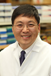
Yutaka Niihara
David Geffen School of Medicine - UCLA , USA
Title: A Phase 3 Study of L Glutamine Therapy for Sickle Cell Anemia and Sickle ß0-Thalassemia
Time : 09:30-09:55
Biography:
Dr. Niihara has been involved in patient care and research for sickle cell disease for most of his career, and is the principal inventor of the patented L-glutamine therapy for treatment of sickle cell disease. He is a cofounder of Emmaus, principal investigator for LABioMed at Harbor-UCLA Medical Center and Professor of Medicine at the David Geffen School of Medicine at UCLA. His experience includes serving as President, Chief Executive Officer and Medical Director of Hope International Hospice. A board-certified by the American Board of Internal Medicine and the American Board of Internal Medicine/Hematology, Dr. Niihara is licensed to practice medicine in both the U.S. and Japan. His honors include the Life Time Achievement Award, from the Sickle Cell Disease Foundation of California and the Abigail Kawananako Award. Dr. Niihara received his B.A. in Religion from Loma Linda University and obtained his M.D. degree from the Loma Linda University School of Medicine.
Abstract:
Background: New treatments for patients with Sickle Cell Disease (SCD) are needed. Oxidative stress may lead to disturbance of cell membranes, exposure of adhesion molecules and damage to the contents of the sickle Red Blood Cells (sRBC). Nicotinamide Adenine Dinucleotide (NAD) molecules modulate oxidation-reduction in sRBCs. Our previous laboratory work demonstrated enhancement of NAD in sRBC by supplementing a precursor of NAD, L-glutamine. In our Phase 2 clinical study in SCD, oral Prescription Grade L-Glutamine (PGLG) signaled a decreasing trend for painful crises at 24 weeks and a significant decrease in hospitalization at 24 weeks. Methods: A randomized (2:1) Phase 3 placebo-controlled trial was conducted across the United States. Subjects were stratified by hydroxyurea usage. Eligibility criteria included patients ≥ 5 years of age with diagnoses of HbSS or HbS/β0-thalassemia with at least two episodes of Sickle Cell Crises (SCC) during the 12 months prior to screening. PGLG at 0.6 g/kg/day (max 30 g), or placebo, was self-administered in two divided doses orally. The primary endpoint was number of SCC; secondary endpoints included rates of hospitalization and adverse events; additional analyses included cumulative hospital days, incidence of Acute Chest Syndrome (ACS) and time to first crises. Results: A total of 230 patients were enrolled at 31 sites. Groups were well balanced for clinical characteristics. The median incidence of SCC (3 vs. 4 events; p=0.008) as well as hospitalizations (2 events vs. 3; p=0.005) was significantly lower in the treatment group compared to the placebo group. Median cumulative hospital days were lower by 41% in the treatment group (6.5 days) vs. the placebo group (11 days) (p=0.022); ACS was 11.9 % in the treatment group and 26.9% in the placebo group (p=0.006). The median time to first crisis was 87 days in the treatment group vs. 54 days in placebo group (p=0.010). Analysis by hydroxurea use, age, and gender yielded consistent findings. Adverse events in the treatment arms were similar between groups. Conclusion: This Phase 3 study in SCD demonstrated that treatment with PGLG provided clinical benefit over placebo by reducing the frequency of painful crises and hospitalization. Additional benefit was observed when evaluating ACS, time to first crises and duration of hospital stay. PGLG was relatively easy to administer and did not require special monitoring.
Seung Eun Choi
Healthcare Company, Korea
Title: The concept of recycling translational medicine
Time : 09:55-10:20

Biography:
Seung Eun Choi has completed her MD from College of Medicine, Seoul National University, and surgery residency from Seoul National University Hospital. She completed her clinical fellowship in pediatric surgery in Seoul National University Children Hospital. She has completed her Ph.D in transplant immunology as full-time researcher from Microbiology and Immunology department in College of Medicine, Seoul National University. Previously, she did in-vivo experiment in immunology area and is working in the healthcare company.
Abstract:
The concept and definition of translational medicine has been developed and expanded, according to the different meaning to different stakeholders. The definition and concept used to be around biomedical research itself in the early time and has been developed to public science currently. Consensus for definition is important because it would be followed by funding from investors. In addition, translational medicine has been developed, resulted from the necessity of the practice rather than the need of academic area. More kinds of players are involved in the translational medicine system currently. For example, patients join in the translational medicine as decision makers as well as subjects in clinical trials. Patients’ role as decision maker has been developed in parallel with the development of information technology. More systems have been developed around the T block and phases, in order to get efficiency in the complex system of healthcare field. The T3 has been better established scientifically and well systemized with regulation. Considering the scientific activity of T3, the concept of translational medicine would like to be suggested as “Recycling translational medicine” innovatively, according to the current practice and players for translational medicine in the field.
Youngmi Jung
Pusan National University, Korea
Title: TSG-6 induces liver regeneration by enhancing autophagy in mice with chronic liver damage
Time : 10:20-10:45
Biography:
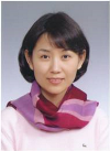
Youngmi Jung has completed her PhD at the age of 32 years from University of Florida College of Medicine and postdoctoral studies from Duke University College of Medicine (division of gastroenterology). She has worked as an assistant professor at Duke University from 2008 to 2010. In 2010, she joined the faculty of the department of Biological Sciences at Pusan National University where she has been working as an associate professor since 2014. Her research is focused on mechanisms underlying the progression of liver diseases and the liver regeneration by stem cells. She has published more than 40 papers in reputed journals.
Abstract:
Tumor necrosis factor-inducible gene 6 protein (TSG-6), one of cytokines secreted from mesenchymal stem/stromal cells, is known to act as an anti-inflammatory factor. Recently, we prove that TSG-6 contributes to the liver regeneration by suppressing the activation of hepatic stellate cells in CCl4-treated mice; however, the mechanism underlying the effect is poorly understood. Autophagy is the catabolic process targeting cell constituents to the lysosomes for degradation and known to be dysregulated in several diseases, including liver diseases. Emerging evidence suggests that the autopagic functions protect hepatocytes from damages. Hence, we hypothesize that TSG-6 promotes the autophagical clearance systems in hepatocytes and protects liver from injury. To prove this hypothesis, mice were fed with Methionine Choline-Deficient Ethionine (MCDE) for 4 weeks, followed by being injected with TSG-6 (TSG-6 group) or saline (MCDE+vehicle group) with MCDE supplemented diets for additional 2 weeks. The histomorphological injuries and increased level of liver enzymes were shown in MCDE-treated mice with or without saline, whereas those observations were markedly ameliorated in TSG-6 group. The markers for autophagosome formation, LC3-II, Lamp2, and Rab7 and autophagy-relate genes, atg3 and atg7, were elevated in TSG-6 group compared to MCDE or MCDE+vehicle group. Immunostaining for LC3, Lamp2 and Rab7 showed that livers of TSG-6 group contained more hepatocytic cells expressing those markers than other groups. Also, Ki67-positive hepatocytic cells were accumulated in TSG-6 group, whereas these cells were rarely detected in MCDE group. Therefore, these results demonstrated that TSG-6 promoted the autophagical function in hepatocytes, contributing to the liver regeneration.
Zijun Zhang
MedStar Union Memorial Hospital, USA
Title: Implantation of autologous adipose tissue-derived mesenchymal stem cells in foot fat pad in rats
Time : 11:05-11:30

Biography:
Zijun Zhang obtained his PhD/MD degree from Military Postgraduate Medical School, Beijing, China and completed his postdoctoral training at University of Melbourne, University of Auckland and the NIH. He is the director of Orthobiologic Laboratory, MedStar Union Memorial Hospital. He has published more than 50 papers in peer-reviewed journals.
Abstract:
The Foot Fat Pad (FFP) bears body weight and may become a source of foot pain during aging. This study investigated the regenerative effects of autologous Adipose Tssue derived Mesenchymal Stem Cells (AT-MSCs) in the FFP of rats. Fat tissue was harvested from a total of 30 male Sprague-Dowley rats for isolation of AT-MSCs. The cells were cultured and induced adipogenic differentiation for one week and labeled with fluorescent dye before injection. AT-MSCs (5x10⁴ in 50µl saline) were injected into the second infradigital pad in right hind foot of the rat of origin. Saline only (50µl) was injected into the corresponding fat pad in the left hind paw of each rat. Rats (n = 10) were euthanized at 1, 2, and 3 weeks and the second infradigital fat pads were dissected for histology. The fluoresence-labeled AT-MSCs were present in the foot fads throughout the 3-week experimental period. On histology, the area of Fat Pad Units (FPU) in the fat pads that received AT-MSC injections was greater than that in the control fat pads. Although the thickness of septae was not changed by AT-MSC injections, the density of elastic fibers in the septae was increased in the fat pads that implanted AT-MSCs. The implanted AT-MSCs largely survived in the weightbearing FFP. There was no evidence that AT-MSCs formed new FPUs, but they stimulated the growth of individul FPUs and formation of elastic fibers in FFP. These data are promising for developing stem cell therapies for aging-associated degeneration in FFP.
Hafiz Ansar Rasul Suleria
The University of Queensland, Australia
Title: Extraction and Characterization of Bioactives from Marine Sources; A Concept of Modern Research and Drug Discovery
Time : 11:30-11:55
Biography:
Abstract:
Marine organisms are increasingly being investigated as sources of bioactive molecules with therapeutic applications as nutraceuticals and pharmaceuticals. Recent trends in functional foods have demonstrated that bioactive molecules play a major therapeutic role in human disease, therefore nutritionists, biomedical scientists and food scientists are working together to discover new bioactive molecules that have increased potency and therapeutic benefits. Marine life constitutes almost 80% of the world biota with thousands of bioactive compounds and secondary metabolites derived from marine invertebrates such as tunicates, sponges, mollusks, sea hares, bryozoans, sea slugs and other marine organisms. These bioactives and secondary metabolites possess antibiotic, anti-parasitic, anti-viral, anti-inflammatory, anti-fibrotic and anti-cancer activities. They are inhibitors or activators of critical enzymes, agonists or inhibitors of transcription factors, competitors of transporters and sequestrants to modulate various physiological pathways. The current review summarises the widely available marine-based nutraceuticals and recent findings, mainly focusing on mode of action, efficacy and underlying mechanisms. It also presents recent research involving the isolation, identification and characterization of marine-derived bioactives with various therapeutic potentials.
Pedro P Medina
University of Granada, Spain
Title: MicroRNAs, Chromatin remodeling complexes and cancer
Biography:
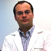
Pedro P. Medina completed his PhD in the Spanish National Cancer Center (CNIO, Madrid Spain) and 5-years of postdoctoral studies Yale University (New Have, CT, USA). He is the Director of the Gene Expression Regulation and Cancer group and Associate Professor at the University of Granada. Pedro has published articles in the field of Cancer more in reputed journals including Nature, Cancer Cell, Nature Methods, Journal of Clinical Oncology, Cell Reports, Oncogene, Human Molecular Genetics, etc.
Abstract:
SWI/SNF chromatin-remodeling complex, which alters the interactions between DNA and histones and modifies the availability of the DNA for transcription. The latest deep sequencing of tumor genomes has reinforced the important and ubiquitous tumor suppressor role of the SWI/SNF complex in cancer. However, although SWI/SNF complex plays a key role in gene expression, the regulation of this complex itself is poorly understood. SMARCA4 is the catalytic subunit of the SWI/SNF complex and it has chromatin-remodeling activity by itself. Significantly, an understanding of the regulation of SMARCA4 expression has gained in importance due to recent proposals incorporating it in therapeutic strategies that use synthetic lethal interactions involving several SWI/SNF subunits. We have displayed that the loss of expression of SMARCA4 observed in some primary lung tumors, whose mechanism was largely unknown, can be explained, at least partially by the activity of microRNAs (miRNAs). Our results show that SMARCA4 expression is regulated by miR-101, miR-199 and especially miR-155 through their binding to two alternative 3'UTRs. These experiments suggest that the oncogenic properties of miR-155 in lung cancer can be largely explained by its role inhibiting SMARCA4. This functional relationship could explain the poor prognosis displayed by patients that independently have high miR-155 and low SMARCA4 expression levels. In addition, these results could lead to application of incipient miRNA technology to the aforementioned synthetic lethal therapeutic strategies.
Sergey Suchkov
I.M.Sechenov First Moscow State Medical University, Russia
Title: Translational ideology and armamentarium as a strategic tool for advancing T1D-related care: Fundamental, applied and affiliated issues

Biography:
Sergey Suchkov was born in 11.01.1957, a researcher-immunologist, a clinician, graduated from Astrakhan State Medical University, Russia, in 1980. He has been trained at the Institute for Medical Enzymology, The USSR Academy of Medical Sciences,National Center for Immunology (Russia), NIH, Bethesda, USA) and British Society for Immunology to cover 4 British university facilities. Since 2005, he has been working as Faculty Professor of I.M. Sechenov First Moscow State Medical University and Of A.I.Evdokimov Moscow State Medical & Dental University. From 2007, he is the First Vice-President and Dean of the School of PPPM Politics and Management of the University of World Politics and Law.In 1991-1995, He was a Scientific Secretary-in-Chief of the Editorial Board of the International Journal “Biomedical Science†(Russian Academy of Sciences and Royal Society of Chemistry, UK) and The International Publishing Bureau at the Presidium of the Russian Academy of Sciences. In 1995-2005, he was a Director of the Russian-American Program in Immunology of the Eye Diseases. He is a member of EPMA (European Association of Predictive, Preventive and Personalized Medicine, Brussels-Bonn), a member of the NY Academy of Sciences, a member of the Editorial Boards for Open Journal of Immunology and others. He is known as an author of the Concept of post-infectious clinical and immunological syndrome, co-author of a concept of abzymes and their impact into the pathogenesis of auto immunity conditions, and as one of the pioneers in promoting the Concept of PPPM into a practical branch of health services.
Abstract:
Type One diabetes (T1D) whilst being a chronic disease, the defining feature of which is the destruction of the insulin-secreting beta-cells and subsequent dependence on exogenous insulin, is possessing with hallmark characteristics of the complex T cell-mediated autoimmunity superimposed on genetic susceptibility. Both genetic and environmental factors combine to precipitate disease, and the outcome of the pathological process is dependent on multiple interrelated factors. The annual global incidence of T1D is increasing by 3-5% per year. Both HLA-genes and immune markers (autoantibodies/autoAbs) have been validated as predictive biomarkers of the subsequent development of the disease in higher-risk relatives and the lower-risk general population. Over the last three decades whilst securing translation of the outcome of basic research in the daily clinical practice using a combination of genomic, proteomic, and metabolomic biomarkers, clinicians are now able to quantify an individual's disease risk from 1 in 100,000 to more than 1 in 2. The HLA genes are reported to account for approximately 40-50% of the familial aggregation of T1D. Current approaches for the prediction of T1D in screening studies take advantage of genotyping HLA-DR and HLA-DQ loci, which is then combined with family history and screening for autoAbs directed against islet-cell antigens. Inclusion of additional moderate HLA risk haplotypes may help identify the majority of children withT1D before the onset of the disease. Fine mapping and functional studies are gradually revealing the complex mechanisms whereby immune self-tolerance is lost, involving multiple aspects of adaptive immunity. The triggering of these events by dysregulation of the innate immune system has also been implicated by genetic evidence. Finally, genetic prediction of T1D risk is showing promise of use for preventives trategies. Understanding of the genetics, environmental factors, and natural history of T1D has led to greater understanding of the etiology and epidemiology of T1D. Accurate prediction is vital for disease prevention so that therapy can be given to those individuals who are most likely to develop the disease. The utility of predictive markers is dependent on three parameters, which must be carefully assessed: sensitivity, specificity and positive predictive value. Specificity is important if a biomarker is to be used to identify individuals either for counseling or for preventive therapy. However, a reciprocal relationship exists between sensitivity and specificity. Thus, successful biomarkers will be highly specific without sacrificing sensitivity. Antiislet autoAbs can be detected before and at time of clinical diagnosis of disease. Autoantibodies (autoAbs) to beta cell antigens (Ags) are used in the diagnosis of T1D, and studies have shown that they can be used to predict risk of developing T1D in first degree relatives of probands. Strategies for disease intervention, therefore, will not only require the induction of T-cell tolerance by tipping the balance towards regulation but will also need to contain approaches that result in the scavenging of inflammatory mediators, in order to facilitate repair. FoxP3-expressing CD4(+) regulatory T cells (Tregs) are potential candidates to control autoimmunity because they play a central role in maintaining self-tolerance. Ultimately, given that clinical diabetes presents late in the autoimmune process, strategies for β-cell regeneration must now be addressed as well. The optimal therapeutic approach for T1D should ideally preserve the remaining β-cells, restore β-cell function, and protect the replaced insulin-producing cells from autoimmunity. SCs possess immunological and regenerative properties that could be harnessed to improve the treatment of T1D; indeed, SCs may reestablish peripheral tolerance toward β-cells through reshaping of the immune response and inhibition of autoreactive T-cell function. There is thus a requirement for an increased, collaborative effort between stem cell biologists and immunologists in order to tailor an optimal therapeutic strategy for the treatment of this debilitating disease whilst translating the basic outcome into the daily clinical practice. There is an immediate need to restore both β-cell function and immune tolerance to control disease progression and ultimately cure T1D.
Fozia Noor
Saarland University, Germany
Title: Advanced human in-vitro models for the study of drug induced cholestasis

Biography:
Fozia Noor did her PhD in Heidelberg, Germany at the Institute of Pharmacy and Molecular Biotechnology in 2006. Currently, she is the group leader of systems biology of mammalian cells and systems toxicology at the Biochemical Engineering Institute of Saarland University. Her research interests include development and optimization of in vitro methods for toxicological screening and metabolomics. Her work has been published in reputed journals.
Abstract:
Cholestasis is among the most common adverse effect leading to drug induced liver injury. Drugs usually disrupt bile acid homeostasis by interfering with their metabolism and transport. Although animals especially rodents have been extensively used in cholestasis research, the clinical translation of this knowledge has been very disappointing. Major reasons are species-specific differences in bile acid composition, metabolism and transport. The purpose of this study was to evaluate the suitability of human HepaRG cells comprising of hepatocytes and cholangiocytes as in-vitro model for screening for cholestatic liver injury. We studied the effects of bosentan which impairs the canalicular excretion of bile acids from the hepatocytes. We also investigated the effects of hydrocortisone as inducer of bile acid metabolism and inhibitor of OATP. Bile acids were analysed by LC-MS. Supplementation of serum concentrations of major bile acids resulted in intracellular accumulation of chenodeoxycholic acid upon Bosentan exposure and glycochenodeoxycholic acid upon hydrocortisone exposure in HepaRG cells. We also investigated the effects of bosentan in 2D and 3D HepaRG cultures for 14 days. Using metabolomics we investigated metabolic and cellular alterations. Repeated dose bosentan exposure resulted in more than 20 fold decrease in the EC50 value. We show that there is a change in cellular metabolism upon 14-day bosentan exposure with metabolome hinting at subcellular changes to adapt to cell stress. In conclusion, such human in vitro models will not only be in valuable in the screening of compounds with cholestatic potential but also in the understanding of the disease.
Sharon Prince
University of Cape Town, South Africa
Title: The binuclear palladacycle, AJ-5, is a promising chemotherapeutic agent in a subset of breast cancers, advanced melanomas and sarcomas

Biography:
Sharon Prince is a Professor in the Department of Human Biology in the Health Sciences Faculty at the University of Cape Town (UCT). She completed a PhD in Cell Biology at UCT and was a Welcome Trust funded postdoctoral fellow at the Marie-Curie Research Institute in the United Kingdom. She heads a cancer biology laboratory and collaborates with leading international researchers, publishes in top journals in the field and has been invited to give lectures at national and international meetings. She was appointed the Director of a breast cancer consortium and is a regular reviewer for journals and funding bodies.
Abstract:
Cancer continues to represent one of the most serious health problems worldwide and there is an urgent need to develop improved anti-tumour therapies. Compared to organic chemotherapeutic molecules, metal complexes offer a much more diverse chemistry and have been shown to have important chemotherapeutic applications. This study describes the anti-tumour activity of a novel binuclear palladacycle complex (AJ-5) in several types of cancers. Compared to normal control cells, AJ-5 is more cytotoxic in a subset of advanced melanoma cells, metastatic breast cancer cells and sarcomas, with IC50 values of less than or equal to 0.20 μM. AJ-5 was found to induce apoptosis by flow cytometry (sub G-1 peak), Annexin V-FITC/propidium iodide double-staining, nuclear fragmentation and an increase in the levels of PARP cleavage. Furthermore, AJ-5 was shown to induce both intrinsic and extrinsic apoptotic pathways as measured by PUMA, Bax, cytochrome c release and active caspases. Interestingly, AJ-5 treatment also simultaneously induced the formation of autophagosomes and led to an increase in the autophagy markers LC3II and Beclin1. Inhibition of autophagy reduced AJ-5 cytotoxicity suggesting that AJ-5 induced autophagy was a cell death and not cell survival mechanism. Moreover, it was shown that AJ-5 induces the ATM-CHK2 DNA damage pathway and that its anti-tumour function is mediated by MAPK signalling pathways. Importantly, AJ-5 treatment efficiently reduced tumour growth in melanoma bearing mice and induced high levels of autophagy and apoptosis markers. Together these findings suggest that AJ-5 may be an effective chemotherapeutic drug in the treatment of several cancers.
Simone Renner
Molecular Animal Breeding and Biotechnology, Germany
Title: Genetically engineered pig models for translational diabetes research

Biography:
Simone Renner has completed her doctoral thesis in 2008 from the Ludwig-Maximilians-University Munich and began her postdoctoral studies at the Chair for Molecular Animal Breeding and Biotechnology of the LMU Munich at that time. She is interested in the establishment and characterization of clinically relevant, genetically-modified pig models for translational diabetes research with the aim to provide large animal models that are predictive for the human situation especially in terms of effect and safety of novel therapeutic approached. Simone Renner has published in reputed journals within the diabetes field and is a member of the DDG and ADA.
Abstract:
For the development and evaluation of novel diagnostic and therapeutic approaches convincing animal models are of essential importance. Rodents are the model of choice for basic biomedical research. However, for the translation into clinical application additional model systems closer to humans are needed. For various reasons the pig seems to be a very suitable model organism: i) as monogastric omnivore it shares many anatomical and physiological similarities with humans, ii) pigs have a favorable reproductive biology, iii) the whole pig genome sequence is available and iv) methods for targeted genetic modification of the pig are well established nowadays. For translational diabetes research we have established different genetically modified pig models. Transgenic pigs expressing a dominant-negative receptor for the incretin hormone Glucose-dependent Insulinotropic Polypeptide (GIP) reveal a reduced incretin effect, reduced glucose tolerance and insulin secretion as well as reduced beta-cell mass and therefore represent important aspects of the prediabetic state. This model was already successfully used for the search of biomarkers during the prediabetic phase and as well as for the characterization of effects of the GLP-1 receptor agonist liraglutide in adolescence. In contrast pigs expressing the mutant insulin C94Y develop hyperglycemia within the first days of life and represent a model for permanent neonatal diabetes. A biobank representing long-standing, poorly controlled diabetes was successfully established in this model. In general these pig models are valuable for numerous applications, e.g. the preclinical evaluation of novel therapeutic approaches, establishment of in vivo imaging techniques, biomarker discovery, regenerative medicine and developmental biology.
Liwei Xie
University of Georgia, USA
Title: Transcriptomic analysis of rongchang pig brain and liver
Biography:
Abstract:
Pig is a one of widely used large animal models in biomedical and translational research as it shares similar characteristics in physiologic and anatomic levels, involving but not limited to cardiovascular and central nervous system. The fast development of Next Generation Sequencing (NGS) provides a useful tool to identify differentially regulated genes in large animals from the human disease-Ââ€relevant aspect. In current investigation, we sequenced RNAs from two major organs in pig, brain, the central nervous operator, and liver, the metabolic process center. From this study, we uncovered the major characteristics of the transcriptional profiles of neuron system-Ââ€related genes in two different organs. Furthermore, other than protein coding genes in two organ, we also identified ~2.5% noncoding genes, including microRNA, and long noncoding RNAs, which may paly an essential regulatory role in mRNA function. The results from current study provide a spectrum of gene expression profile information that can be further applied to the biomedical and clinical investigation of neurological and metabolic disorders related
Ridham Khanderia
Pandit Deendayal Upadhayay Medical College, India
Title: Evaluating the role of indirect bilirubin and urobilinogen as an alternative screening tool for beta thalassemia minor

Biography:
RidhamKhanderia is a final year MBBS , PanditDeendayalUpadhyay Medical College , Rajkot , Gujarat , India. He has done a research project on above topic under ICMR-STS( Indian Council of Medical Research – Short term studentship) program which is under a review process in the reputed journal ( Indian Journal of Medical Research). He has been selected as an Elsevier Student Ambassador-2014. He has been the Vice-President of College Alumni Association for the year 2012-13. He has given Oral Presentation on above topic in “MEDICON†research conference in India. He has secured First Rank in College Final Exams for last three years.
Abstract:
Background & Objectives: Beta thalassemia continues to be significant burden to Western India particularly Saurashtra region of Gujarat.The cost of ideal treatment of 1 thalassemia major child is nearly Rs 1,00000/annum.So emphasis must be shifted from treatment to prevention that includes mass screening.Most of these tests have limited availability and require sophisticated equipments.Thus,there is need for simple,low cost and reliable test.The present study will evaluate validity of such test Indirect Bilirubin and Urine Urobilinogen. Methods: The present crossectional study was conducted on 100 subjects in PDU Medical College Rajkot,Gujarat over period of two months.In first group,50 Beta Thalassemia minor patients were selected while in second group 50 normal individuals from hospital staff were selected.Complete-haemogram,serum-direct,indirect and total bilirubin,urine-urobilinogen and HbA2 levels by electrophoresis were done and results analyzed. Results: Of the 50 cases in first group,41 had higher indirect bilirubin level(>0.7 mg/dl),35 had high urine urobilinogen level(>1 mg/dl),49 had lower(<1530)Shine & Lal index while all 50 had HbA2 level >3.5%.In second group,3 had high bilirubin levels,4 had high urobilinogen levels,only 2 had low(<1530)Shine & Lal-index while no on e had HbA2 level >3.5%.Indirect-bilirubin showed a sensitivity of 82%,specificity of 94%.Urobilinogen showed sensitivity of 70% & specificity of 92%. Shine & Lal index showed sensitivity of 98% & specificity of 96%. Combined Sensitivity & Specificity of bilirubin & urobilinogen was found to be 94% & 98% respectively. Interpretation & Conclusion: Thus,serum-indirect-bilirubin and urine-urobilinogen is a valuable,cost-effective screening test with sensitivity & specificity comparable to RBC indices and so may be used in rural settings that are devoid of automated-cell-counters.


