Day :
- Track-4 Translational Therapeutics and Technologies
Track-5 Translational Modelling of Efficacy and Safety
Track-8 Clinical and Translational Oncology
Track-9 Immunology and Infectious Diseases
Session Introduction
Sergey Suchkov
A.I.Evdokimov Moscow State University of Medicine and Dentistry, Russia
Title: Antibody-proteases as promising tools of novel generation to be translated into clinical practice for having multistep (subclinical and clinical) demyelination monitoring secured
Time : 11:10-11:35

Biography:
Sergey Suchkov was born in 11.01.1957, a researcher-immunologist, a clinician, graduated from Astrakhan State Medical University, Russia, in 1980. He has been trained at the Institute for Medical Enzymology, The USSR Academy of Medical Sciences,National Center for Immunology (Russia), NIH, Bethesda, USA) and British Society for Immunology to cover 4 British university facilities. Since 2005, he has been working as Faculty Professor of I.M. Sechenov First Moscow State Medical University and Of A.I.Evdokimov Moscow State Medical & Dental University. From 2007, he is the First Vice-President and Dean of the School of PPPM Politics and Management of the University of World Politics and Law.In 1991-1995, He was a Scientific Secretary-in-Chief of the Editorial Board of the International Journal “Biomedical Science” (Russian Academy of Sciences and Royal Society of Chemistry, UK) and The International Publishing Bureau at the Presidium of the Russian Academy of Sciences. In 1995-2005, he was a Director of the Russian-American Program in Immunology of the Eye Diseases. He is a member of EPMA (European Association of Predictive, Preventive and Personalized Medicine, Brussels-Bonn), a member of the NY Academy of Sciences, a member of the Editorial Boards for Open Journal of Immunology and others. He is known as an author of the Concept of post-infectious clinical and immunological syndrome, co-author of a concept of abzymes and their impact into the pathogenesis of auto immunity conditions, and as one of the pioneers in promoting the Concept of PPPM into a practical branch of health services.
Abstract:
Among the best-validated proteome-related predictive biomarkers, antibodies (Abs) are the best known. A combination of different panels of autoAbs in the diagnostic practice is becoming of great significance to predict risks of chronization of the autoimmune disorder since most of the latter are preceded by a long subclinical (symptom-free) phase when the patients or persons-at-risk could be identified via specific sets of autoAbs. Of particular interest would be algorithms which would employ panels of targeted Abs for screening patients and their relatives at risks for the presence of preclinical (lab proof-based) signs and for thus having the data translated into the daily practice. Abs against myelin basic protein/MBP endowing with targeted and highly specific proteolytic activity (Ab-proteases) are appearing to be of great value to monitor demyelination at either of the stages. The activity of Ab-proteases identified and purified from MS blood samples markedly differed between: (i) MS patients and healthy controls; (ii) different MS courses; (iii) EDSS scales of demyelination to correlate with the disability of MS patients to predict transformations prior to changes of the clinical course. The sequence-specificity of Ab-proteases demonstrates five sites of preferential proteolysis to be located within the immunodominant region of MBP. The activity of Ab-proteases was first registered at the subclinical stages. About 24% of the direct MS-related relatives were seropositive for low-active Ab-proteases from which 50% of the seropositive relatives monitored for 2 years have been demonstrating stable growth of the proteolytic activity. And, finally, primary clinical manifestations observed were coincided with the activity to have its mid-level reahced. The activity of Ab-proteases in combination with their sequence-specificity to attack well-defined epitopes to be released from MBP during epitope spreading, would confirm a practical value of Ab-proteases demonstrating their unique functionality. Meanwhile, Ab-proteases can be programmed and reprogrammed to suit complex cell biochemistry. And thus two logical questions would arise: (i) would the original potential for the Ab-mediated proteolysis relate to natural Ab-related function? (ii) could that potential be translated into the clinical practice to suit the need of clinicians? Canonical Abs play neither predictive nor discriminative role to affect the subclinical stage of MS. Meanwhile, sequence-specific Ab-proteases have proved to be greatly informative and thus valuable as translational biomarkers to monitor MS at both subclinical and clinical stages. So, the activity in combination with the sequence-specificity would confirm a high subclinical and predictive value of Ab-proteases as applicable for personalized monitoring protocols. Moreover, of tremendous value in this sense are Ab-proteases directly affecting the physiologic remodeling of tissues with multilevel architectonics (for instance, myelin). So, targeted Ab-mediated proteolysis could be also applied to isolate from Ig molecules catalytic domains directed against encephalitogenic epitopes or domains containing segments to exert proteolytic activity. And further studies on Ab-mediated MBP degradation and other targeted Ab-mediated proteolysis may provide biomarkers of new generations and thus a supplementary tool for assessing the disease progression and predicting disability of the patients and persons-at-risks. The latter means that translation of the data collected on targeted Ab-mediated proteolysis may provide a supplementary tool for predicting demyelination and thus the disability of the MS patients.
Isabelle De Waziers
INSERM U1147, France
Title: Use of engineered mesenchymal stem cells against cancers
Time : 11:35-12:00

Biography:
She has completed her Ph.d in Molecular Pharmacology, Experimental Pharmacology and Metabolism, Physiology of Nutrition. She is the member of administration council of the Faculty of Medicine in Paris Descartes University, member of the American Association for Cancer Research. She has been appointed as the Mayor of Lignières en Vimeu from 1995, President of the Community of Communes of the Region of Oisemont from 2014,Departemental councilor from 2014. Her special area or research are Xenobiotic metabolism, Toxicology, Molecular Biology, Cancer, Resistance to chemotherapy, Gene therapy, Transcriptomic, Metabonomic.
Abstract:
Gene-Directed Enzyme Pro-Drug Therapy (GDEPT) consists in expression of a suicide gene in tumor cells allowing in situ conversion of the pro-drug into cytotoxic metabolites. In a previous work, we demonstrated that the combination of a mutant CYP2B6 with NADPH Cytochrome P450 reductase (CYP2B6TM-RED), as a suicide gene, and Cyclophosphamide (CPA) might constitute a powerful treatment for solid tumors (Touati et al, Curr Gene Ther, 2014). The efficiency of this combination was mainly due to i) an optimized suicide gene able to metabolize efficiently CPA ii) an efficient bystander effect, iii) the development of an antitumor immune response. Major impediments were the targeting of the suicide gene specifically to the tumor cells and its low expression into tumor cells. Recently, therapies based on genetically engineered Mesenchymal Stem Cells (MSCs) expressing suicide gene have received a great deal of attention because of their therapeutic potential to treat solid tumors (Kosaka et al, Cancer Gene Ther, 2012). Indeed, MSCs possess an extraordinary ability to home into tumors due to the inflammatory mediators which are found at the site of a tumor. MSCs can be easily isolated, from tissues such as bone marrow (BM-MSCs) and adipose tissue (ACS), expanded in culture and efficiently transduced with recombinant viral vectors leading to important and stable expression of the suicide gene. Once the transduction is performed, the most efficient clone for bioactivation of the prodrug can be selected and used for several patients. Indeed, given the minimal expression of the major histocompatibility complex MHC-I and MHC-II, allogeneic expanded Adipose-derived Stem Cells (eASC) delivered locally are well-tolerated and currently in clinical Phase III clinical trials for the treatment of inflammatory and auto-immune diseases (www.tigenix.com). Murine MSCs were genetically engineered ex-vivo to stably express luciferase or CYP2B6TM-RED. The most efficient clones were selected. MSCs expressing luciferase were used to follow the future of MSCs after their intratumoral injections in animal models. MSCs expressing CYP2B6TM-RED were used to check ex vivo and in vivo their efficiency to bioactivate CPA and destroy neighboring tumor cells through a by-stander effect. Intratumoral injections of CYP2B6TM-RED-MSCs and CPA allowed a complete eradication of the tumor in 33% of the mice without any recurrence four months later. Different experiments are now under investigation to improve the efficiency of this strategy.
Sevtap Savas
University of Newfondland, Canada
Title: Identification of genetic variations associated with outcome risk in colorectal cancer: Where are we now?
Time : 12:00-12:25

Biography:
Sevtap Savas obtained her PhD in Molecular Biology and Genetics from the Bogazici University, Turkey. She trained as a post-doctoral fellow or research associate in Louisiana State University (USA), Mount Sinai Hospital Research Institute (Canada) and Princess Margaret Hospital/Ontario Cancer Institute (Canada). Since 2008, she has been a faculty member at Discipline of Genetics, Memorial University of Newfoundland (Canada). Her research program focuses on genetic prognostic studies in colorectal cancer using genetic, epidemiological, biostatistical, and computational approaches as well as development of public databases. She also serves as a reviewer, academic editor or editorial board member for several journals.
Abstract:
My laboratory is interested in identifying genetic markers that can help predict the patient outcomes in colorectal cancer. For this purpose, associations of genetic variations, such as SNPs (Single Nucleotide Polymorphisms) and CNVs (Copy Number Variations), with patient survival times are investigated using statistical methods. In collaboration with the investigators at the Newfoundland Colorectal Cancer Registry (NFCCR) and other institutions, my laboratory conducted a number of candidate gene, candidate pathway, and genome wide prognostic studies in colorectal cancer. In this presentation, results of select projects will be discussed.
Jan Scicinski
EpicentRx Inc , Mexico
Title: RRx-001, a novel ROS-mediated epigenetic modulator: ‘Episensitization’ to previously failed therapies
Time : 12:25-12:50 PM

Biography:
Dr Scicinski has 30 years experience in the pharmaceutical and biotech industries in drug discovery and development. He is Senior VP & Chief Scientific Officer at EpicentRx, Inc leading drug discovery, basic research and preclinical development efforts. He oversees regulatory affairs, CMC and QA. Prior to EpicentRx, Dr Scicinski held leadership positions in research on drug delivery and discovery at DURECT and ALZA and in drug discovery and chemical technology at Nuada and GSK. Dr Scicinski holds a BSc from Imperial College, London and a PhD from Cambridge University and has co-authored over 90 peer-reviewed publications, abstracts and book chapters.
Abstract:
Acquired resistance to chemotherapies in cancer results in early disease progression that translates to lower survival. Resensitization of tumors to previously effective but now failed therapies is an established paradigm in ovarian cancer, which could be expanded to include other solid tumors, such as colorectal, NSCLC, SCLC and cholangiocarcinoma, leading to across-the-board improvements in patient survival and radiologic response. Although epigenetic agents hold the promise of priming tumors to subsequent chemotherapy, many agents are too toxic for chronic use. RRx-001, a novel ROS-mediated epigenetic modulator with activity against DNMTs and HDACs, is systemically non-toxic, with improvements in QOL observed in clinical trials making it an ideal candidate for ‘episensitization’ applications. RRx-001 resensitization was observed in Phase 1 and is being investigated in multiple Phase 2 studies: “ROCKET” is a two stage clinical study in metastatic colorectal cancer investigating resensitization to irinotecan-based therapies. Patients who previously responded, then progressed on irinotecan-based therapies are randomized to receive RRx-001 (1x week) or regorafenib (target ~190 pts) until progression or unacceptable toxicity (Stage 1). Patients then receive irinotecan-based therapies (Stage 2). The primary endpoint is overall survival with secondary endpoints to investigate resensitization. The trial is continuing. RRx-001 patients entering Stage 2 are showing improving survival, marked CEA decreases and radiological responses. In contrast, regorafenib patients eligible for stage 2 were too systemically unwell to proceed. Early results suggest that RRx-001 may resensitize patients to irinotecan-based therapies and appear to be generalized, potentially translating to increased overall survival.
Eli Raveh
The Hebrew University of Jerusalem, Israel
Title: From chromosomal instability to metastases- H19’s role in cancer progression and its clinical significance
Time : 13:50-14:15
Biography:
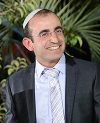
Eli Raveh earned his PhD in developmental biology from the Hebrew University of Jerusalem (HUJI) in 2011, specializing in hair and skin development. Since then he serves as a research assistant in Prof. Avraham Hochberg’s Laboratory,at the department of biological chemistry, HUJI, which focuses on cancer therapy research based on the H19 gene involvement in cancer.
Abstract:
The oncofetal H19 long non-coding RNA (lncRNA) is normally expressed in the embryo and downregulated at birth; however its expression is re-elicited in Human tumors of almost every type. Accumulating data by us and others consolidate into a paradigm according to which H19 is in the center of a mammalian “selfish survival pathway” activated by the cancerous cell in order to cope with stress conditions. P53 nullification or dysfunction, hypoxia, serum starvation, chemotherapy and other stress inducers upregulate H19 expression. H19 in turn supports tumor growth and enhances proliferation in response to hypoxia and p53 mutations. We have also recently proven that H19 RNA significantly contributes to Epithelial to Mesenchymal Transition (EMT), a process which is known to emerge as a response to a multitude of stress conditions. Surprisingly, studies indicate that H19 is involved also in the further, apparently converse steps of mesenchymal to epithelial transition to support colonization and proliferation in the secondary tumor site. In view of the paradigm suggested above, H19’s unique expression in cancers, and the active involvement of H19 in almost every deleterious aspect of cancer progression, H19 should serve as a state of the art target for highly selective cancer therapy. Indeed, we currently use a DNA-plasmid based drug that harnesses H19 regulatory elements to drive the expression of Diphtheria toxin - a translational regulated therapy designed to selectively kill cancer cells with no “collateral damage” to the healthy surrounding tissues. We will comprehensively review recent studies by us and others that align with our paradigm, and demonstrate how understanding of H19 expression and biology is translated into promising results in bladder, pancreas and ovarian cancer clinical trials.
Suoqin Tang
PLA General Hospital, China
Title: Survivin siRNA nano particles are capable of inhibiting cancer cell growth both in vitro and in vivo
Time : 14:15-14:40

Biography:
Suoqin Tang has completed his MD at the age of 23 years from The Fourth Medical University in China and postdoctoral studies from University of Southern California School of Medicine. He is chief physician and professor of Department of Pediatrics, Chinese PLA General Hospital, a famous and one of the best hospitals in China. He has published more than 65 papers both in China and overseas. He is an international member of Childrens Oncology Group(US), standing member of Chinese Pediatric Society, and editor of Chinese Journal of Pediatrics. He is doing clinical work on chemotherapy of leukemia and solid tumors, including lymphoma, neuroblastoma and PNET, his research work focus on target therapy of cancer.
Abstract:
Since the stability of siRNA molecules in the blood and efficiency of siRNA delivery into target organs or tissues following systemic administration have been the major issues that limit applications of siRNA in human patients, we try to explore if siRNA liposome entrapment works in the development of novel therapeutics. Our study aims to evaluate the therapeutic effect of survivin siRNA nano particles, on liver cancer, colon cancer and cervical cancer both in vitro and in vivo. First, sequences of survivin siRNA we designed had been screened for their efficacy, and the most effective one was chosen for the next study. Second, we have tested the biological effect of survivin siRNA nanoparticles on cancer cell lines in vitro, and resulted that, the survivin mRNA and its protein expression was significantly inhibited in MHCC-97H cells, HeLa cells and LoVo cells, also proliferation of those cell lines was inhibited and apoptosis was promoted. Third, subcutaneous xenograft Balb/c nude mice of MHCC-97H cells, HeLa cells or LoVo cells were established. Survivin siRNA nanoparticles(70.7±29.077nm in size), with dosage of 3 mg/kg survivin siRNA was given via local or intravenous injection, twice a week continuously for 4 weeks. As results, significant tumor growth inhibition both in local injection and venous injection was observed, compared to control mice who received scribbled siRNA nan particles at the same dosage, with inhibition rate of 32.22% and 36.67%, at the end of study in venous injection and local injection respectively, the relative tumor volume in mice with local injection showed significant less than control group from 10 days after first injection((P<0.05), till 31 days, it was also the same situation in mice with venous injection compared to control, (P<0.05) except day 17, survivin mRNA and its protein were down regulated at the end of study, compared to control group. Our study also showed some inhibition activity of survivin siRNA nanoparticles in subcutaneous xenograft Balb/c nude mice of HeLa or LoVo cells, survivin mRNA and protein expression was all down regulated in tumor tissue compared to control. We labeled the survivin siRNA with Cy3 florescence and intravenous injection to nude mice with MHCC-97H cells, and found intense Cy3 distribution in tumor mass, liver and spleen, but scant distribution in heart, brain, lung, bone marrow and gastrointestinal tract. Our study revealed that survivin siRNA nanoparticles were capable of inhibiting tumor growth both in vitro and in vivo.
Huiyun Zhang
Liaoning Medical University, China
Title: Subsets of regulatory T cells and their roles in allergy
Time : 14:40-15:05

Biography:
Huiyun Zhang has completed her PhD at the age of 36 years from Shantou University Medical College and postdoctoral studies from MacMaster University School of Medicine. She is the director of pathophysiology Department to Liaoning Medical University. She has published more than 50 research articles including 22 recorded in Scientific Citation Index and has been serving as an editorial board member for several Journals. Her research interest is to explore the diagnostic and therapeutic procedures based on the pathophysiology of allergy. Accordingly, she has obtained 3 National Natural Science Foundation of China (No. 81030054, 81241135, 81472016) and 1 Key Provincial Health Project. And she has participating in 2 Major State Basic Research Program of China (973 Program, No. 2013CB530500&2007CB512400) and 1 national sci-tech support plan (2014BAI07B02) as well as editing “Theoretical Allergology ”, “Experimental Allergology”, “Clinical Allergology”, “Allergology in Ear Nose Throat Head and Neck Diseases” and “Frontier Progress in Immunology” a total of five books on Allergology and Immunology in Chinese
Abstract:
In recent years, it is recognized that acquired immunity is controlled by regulatory T cell (Treg). Since fundamental pathophysiological changes of allergy are mainly caused by hyperresponsiveness of immune system to allergens that acquires after birth, Tregs likely play key roles in the pathogenesis of allergy, particularly during the sensitization phase. However, accumulated information indicate that there are several distinctive subtypes of Tregs in man, and each of them seems to play different role in controlling immune system, which complicates the involvement of Tregs in allergy. The aim of the present study is to attempt to classify subtypes of Tregs and summarize their roles in allergy. Tregs should include natural Tregs (nTreg) including inducible costimulator (ICOS)(+) Tregs, inducible/adaptive Tregs (iTreg), interleukin (IL)-10-producing type 1 Tregs (Tr1 cells), CD8(+) Tregs and IL-17-producing Tregs. These cells share some common features including expression of Foxp3 (except for Tr1 cells), and secretion of inhibitory cytokine IL-10 and/or TGF-ï¢. Furthermore, it is noticeable that Tregs likely contribute to allergic disorders such as dermatitis and airway inflammation, and play a crucial role in the treatment of allergy through their actions on suppression of effector T cells and inhibition of activation of mast cells and basophils. Modulation of functions of Tregs may provide a novel strategy to prevent and treat allergic diseases.
Geeta Shroff
Nutech Mediworld, India
Title: Validating the efficacy of the Human Embryonic Stem Cell (hESC) therapy in cerebral palsy patients

Biography:
A graduate in medicine from the University of Delhi, Dr. Shroff did her post graduation in Gynecology & Obstetrics. She is specialized in treating infertility, and is a trained embryologist and a qualified IVF practitioner. After gaining 8 years of valuable clinical experience at Safdarjung Hospital & Batra Hospital, Dr. Shroff set up her own IVF practice in 1996. She began research on human embryonic stem cells in 1999, and pioneered human embryonic stem cell therapy. Since 2002, Dr. Shroff has treated more than 1300 patients suffering from various conditions-Spinal cord injuries, Diabetes, Multiple sclerosis, Parkinson’s disease, Cardiac conditions, Cerebral palsy.
Abstract:
Introduction: We have previously shown that hESCs have the potential to treat Cerebral Palsy (CP) related neurological impairments. Here, we focus on the validation of efficacy of hESC therapy in patients with CP. Methodology: This was a record based study (October 2007 to July 2013) of 101 CP patients (age <54 years). hESCs were given in four treatment sessions (T1- T4). The variables “ILLNESS” (How ill at start) and “EMENDMENT” (How emended at end of session) were analyzed. Results: Of 101 patients, 73 attended T2, 41 attended T3 and 15 attended T4. For T1, the average duration of therapy (TD 1) was 60 days. At the end of T1, patients had improved by ~180% from baseline. Similarly, for T2, T3 and T4 (TD 2, 3 = 59 days; TD 4 = 60 days), an improvement by 162%, 153% and 170%, respectively from end of previous session was noted. During T1, T2, T3 and T4 the “ILL” patients, who were ‘severe’ (53, 36, 20, and 6) at admission, emended as endured (40, 28, 17, and 5) and profuse (13, 8, 3, and 1) at discharge. Moderate “ILL” patients (48, 48, 2, and 9) at admission emended as “profuse” (36, 28, 17, and 7) and “endured” (12, 9, 4, and 2) at discharge. For severe cases at admission, the odds of profuse emending when therapy ended were 0.11, 0.09, 0.75, and 0.04 times lower than the cases that were moderate at admission. Conclusion: hESCs therapy caused betterment in the condition of CP patients compared to first admission during each session.
Chi-Tai Yeh
Taipei Medical University, Taiwan
Title: Potential clinical application of PG2 (Astragalus polysaccharide) in cancer-related fatigue and immune modulation
Time : 15:50-16:15

Biography:
Chi-Tai Yeh received his Ph.D. in Food Science and Biotechnology from National Chung Hsing University. Dr. Yeh is currently the Research Fellow in the Graduate Institute of Clinical Medicine at the Taipei Medical University. He is also the Deputy Director of Department of Research and Education of Taipei Medical University-Shuang Ho Hospital. Dr. Yeh has contributed 2 book chapters, published 45 articles in the field of cancer & nutritional chemistry journal, and got 5 patents in the medical compound of cancer therapy. His major research interests include in cancer cell biology, cancer stem cell research, nutrigenomis and cancer chemoprevention with dietary phytochemicals.
Abstract:
A major complaint (>90%) amongst cancer patients undergoing chemotherapy is fatigue, and it remains an unmet medical challenge. Cancer-Related Fatigue (CRF) is well defined in the 10th revision of the International Statistical Classification of Diseases and Related Health Problems (ICD-10). Coupled with the limited understanding of the etiological basis of CRF, current treatment options, such as psycho-stimulants and hematopoietic agents, are ineffective. However, there is increasing recognition of the role of inflammatory cytokines, especially IL-6 and TNF-alpha on CRF pathogenesis. . The prevailing model of cancer treatments often results in the dysregulation of the immune system, which in turn negatively modulate the Central Nervous System (CNS). Traditional Chinese Medicine (TCM) has a long history of treating patients who suffer from deficient ‘qi’ and blood, as well as imbalance of ‘yin’ and ‘yang’. Interestingly, modern science now recognizes the immune modulating activity of many TCM compounds. PG2, a highly purified polysaccharide portion of Astragalus membranaceus, activates immune cells and influences the cytokine network. A pivotal randomized, double-blind, placebo-controlled clinical trial investigated the therapeutic efficacy of PG2 for CRF treatment in advance cancer patients, and found that PG2 significantly alleviated CRF: 82% of the patients had reduced fatigue symptoms after the first PG2 cycle. Thus, PG2 appears to be an effective treatment for CRF. For the indication of CRF in palliative care, PG2 was TFDA-approved for commercial distribution in Taiwan since 2010. The present study focuses on PG2’s immune-modulating function, such as its effect on inflammation and the tumor microenvironment. Tumor-Associated Macrophage (TAM) plays a crucial role in tumor growth, progression and metastasis, and is a target for TCMs such as PG2. Furthermore, we have established an algorithm for early detection of CRF and its accurate diagnosis, based on the correlation of several cytokine biomarkers and CRF questionnaires.. In conclusion, it is anticipated that our work will further shatter the myth of the therapeutic intractability of CRF and push the limits of integrative medical care.

Biography:
Wei-Dong Han, MD, PhD, is the Director of Department of Molecular Immunology/Bio-therapeutic, Director of Department of Stem Cell and Tissue Regeneration in Chinese PLA General Hospital. He is a pioneer in the field of tumor immunotherapy, initially developed the clinical translation of chimeric antigen receptor T (CART) cells in China. He holds 10 projects of clinical trial, including 8 registered CART-based trials (CART19, CART20, CART30, CART33, CART-EGFR, CART-HER-2, and CART-138). The corresponding patents were also applied or obtained in China. In recent 10 years, he obtained 9 grants in China and published more than 80 papers in international journals.
Abstract:
Patients with relapsed or refractory Non-Hodgkin lymphoma (NHL) have a dismal prognosis. Chimeric antigen receptor (CAR) modified T cells targeting CD20 have shown activity in phase I clinical trial for patients with advanced B-cell lymphomas. We performed a phase IIa trial to further assess the safety and efficacy of administering autologous anti-CD20 CAR T (CART-20) cells to patients with refractory or relapsed CD20+ B-cell lymphoma. Eleven patients including one with previous auto-HSCT and one primary cutaneous B-cell lymphomas were enrolled. Seven patients underwent cytoreductive chemotherapy for tumor debulking and lymphocyte depletion before T cell infusions. CART-20 cells were infused into patients at doses of 2.8 to 14.6×10⁶ cells/kg. The overall objective response rate was 9 of 11 (81%), with 6 complete remissions (CR) and 3 partial remissions (PR); no severe toxicity was observed. The median PFS had lasted for more than 10 months with 1 maintaining for 25-month continuous CR. A significant inverse correlation between the molecule levels of the CAR gene and disease recurrence or progression was observed. Additionally, it was deservered to be addressed clinically that the lesions in some special sites such as in spleen and testicle were refractory to CART-20 treatment.
Kuo-Hsiang Chuang
Taipei Medical University, Taiwan
Title: Expressing membrane antibody reporter in the islet cells of nonobese diabetic mice to noninvasive image the disease progression of autoimmune diabetes
Time : 16:40-17:05

Biography:
Dr. Kuo-Hsiang Chuang completed his Ph.D. degree in biomedicine at Kaohsiung Medical University, Taiwan, in 2010. From March 2010 to January 2012, he joined Professor Tian-Lu Cheng’s group (Kaohsiung Medical University) as a Postdoctoral Fellow to study protein engineering, including the development of humanized antibodies and novel recombinant protein drugs. In February 2012, He became an assistant professor in Graduate Institute of Pharmacognosy, Taipei Medical University, Taiwan. Now, he focuses on several research fields, including: reporter genes/noninvasive imaging systems, protein engineering, immunotherapy, and type 1 diabetes.
Abstract:
Expressing a reporter gene in the pancreatic islet of Non-Obese Diabetic mice (NOD mice) allows researchers real-time monitoring the processes of islet loss by noninvasive imaging systems, overcoming the defect of traditional method in which researchers need to weekly sacrifice lots of NOD mice to observe the insulitis in pancreatic islet section.Toward achieving this aim, we previously developed a murine anti-Polyethylene Glycol antibody receptor (anti-PEG reporter) which can be stably expressed on cell surface and can selectively trap PEGylated imaging probes (PEG-NIR797, PEG-124I, and PEG-SPIO) to assess the delivery of cells in vivo by different imaging systems (optical imaging, micro-PET, and MRI). Currently, we have successfully developed the NOD/pIns-αPEG mice which stably express the anti-PEG reporter in their pancreatic islets by using an insulin promoter.The PEG-NIR797 fluorescent probe can specifically accumulate at the pancreatic islet region of NOD/pIns-αPEG mice but not control NOD mice, assisting researchers in conveniently and accurately tracing the process of islet loss and further investigating the islet-protective effects of drugs or genes by optical imaging system.Importantly, expressing the anti-PEG reporter in the pancreatic islet of NOD/pIns-αPEG mice does not affect the islet size, insulin secretion, and the disease progression of type 1 diabetes. The NOD/pIns-αPEG mice may help researchers easily tracing the disease progression of type1 diabetes by noninvasive imaging systems, further providing a valuable tool for worldwide pharmaceutical companies and drug research institutes to screen and evaluate the diabetes drugs.
Solomon Umukoro
University of Ibadan, Nigeria
Title: Mechanisms involved in memory promoting effect of Jobelyn® in mice exposed to unpredictable chronic mild stress

Biography:
Umukoro S has completed his PhD from University of Lagos, Nigeria. He is the Chairman of Central Animal House, University of Ibadan, Ibadan. He has published more than 35 papers in reputed journals and has been serving as a resource person for both local and international journals.
Abstract:
This study was designed to evaluate the possible mechanism (s) involved in memory promoting effect of Jobelyn® in mice exposed to unpredictable chronic mild stress (UCMS). Male Swiss mice were given JB (5-25 mg/kg, p.o) 30 min before exposure to UCMS for 14 days before testing for memory function. Serum corticosterone level was estimated using ELISA kits while the levels of malondialdehyde and glutathione as well as acetylcholine esterase activity in the brain homogenate were determined spectrophotometrically. Histology of the brain tissues and estimation of the populations of viable neurons in the hippocampal regions were done after staining with hematoxyline and eosin. JB reversed memory impairment and also decreased serum corticosterone level produced by UCMS (p<0.05). Moreover, JB reduced malondialdehyde levels and elevated the concentrations of glutathione in the brain of mice exposed to UCMS. Also, JB decreased brain acetylcholine esterase activity when compared with chronic stress group (p<0.05). JB offered a significant protection against UCMS-induced degeneration and death of neuronal cells of the Cornu Ammonis (CA3) of the hippocampus region of the brain suggesting neuro-protection. These findings suggest that Jobelyn® attenuated memory deficits induced by UCMS in mice and may be useful therapeutically for the treatment of stress-related cognitive dysfunctions. The antioxidant, anti-cholinesterase and neuroprotective activities demonstrated by Jobelyn® may be playing a significant role in its memory promoting effect in mice exposed to unpredictable chronic mild stress. The reduction in serum corticosterone, a major indicator of stress response further supports its usefulness in stress-related disorders. The adaptogenic-like activity of MJ may be related to the modulation of serum corticosterone levels, inhibition of anti-choline esterasoxidative stress and neuro-protective effect.
Solomon Umukoro
University of Ibadan, Nigeria
Title: Mechanisms involved in memory promoting effect of Jobelyn® in mice exposed to unpredictable chronic mild stress

Biography:
Umukoro S has completed his PhD from University of Lagos, Nigeria. He is the Chairman of Central Animal House, University of Ibadan, Ibadan. He has published more than 35 papers in reputed journals and has been serving as a resource person for both local and international journals.
Abstract:
This study was designed to evaluate the possible mechanism (s) involved in memory promoting effect of Jobelyn® in mice exposed to unpredictable chronic mild stress (UCMS). Male Swiss mice were given JB (5-25 mg/kg, p.o) 30 min before exposure to UCMS for 14 days before testing for memory function. Serum corticosterone level was estimated using ELISA kits while the levels of malondialdehyde and glutathione as well as acetylcholine esterase activity in the brain homogenate were determined spectrophotometrically. Histology of the brain tissues and estimation of the populations of viable neurons in the hippocampal regions were done after staining with hematoxyline and eosin. JB reversed memory impairment and also decreased serum corticosterone level produced by UCMS (p<0.05). Moreover, JB reduced malondialdehyde levels and elevated the concentrations of glutathione in the brain of mice exposed to UCMS. Also, JB decreased brain acetylcholine esterase activity when compared with chronic stress group (p<0.05). JB offered a significant protection against UCMS-induced degeneration and death of neuronal cells of the Cornu Ammonis (CA3) of the hippocampus region of the brain suggesting neuro-protection. These findings suggest that Jobelyn® attenuated memory deficits induced by UCMS in mice and may be useful therapeutically for the treatment of stress-related cognitive dysfunctions. The antioxidant, anti-cholinesterase and neuroprotective activities demonstrated by Jobelyn® may be playing a significant role in its memory promoting effect in mice exposed to unpredictable chronic mild stress. The reduction in serum corticosterone, a major indicator of stress response further supports its usefulness in stress-related disorders. The adaptogenic-like activity of MJ may be related to the modulation of serum corticosterone levels, inhibition of anti-choline esterasoxidative stress and neuro-protective effect.
Osama Rahma
University of Virginia, USA
Title: Harnessing the immune microenvironment of gastrointestinal cancers using combined modalities

Biography:
Osama Rahma has completed his MD from Damascus University and Internal Medicine Residency from East Carolina University. He completed a fellowship in Immunotherapy and Medical oncology from the Vaccine Branch and the Medical oncology Branch at the National Cancer Institute. He is currently the leader of the hepatobiliary and pancreatic cancer program at University of Virginia. Dr. Rahma is a Principal Investigator on few multicenter clinical trials in pancreatic cancer and has published more than 15 papers in reputed journals.
Abstract:
Immunotherapy has recently emerged as a promising modality in cancer treatment leading to the approval of immune checkpoint inhibitors in few malignancies including melanoma and lung cancer. Tumor-Infiltrating Lymphocytes (TILs) play a major role in anti-tumor immune responses, and their presence is correlated with survival in a variety of tumors. However, These TILs do not reach the Pancreatic Cancer (PC) cells in significant numbers due to the presence of stroma and suppressive microenvironment. In addition, some Colorectal Cancers (CRC) have low immune scores, recently described by Galon et al to assess the presence of TILs in CRC. Furthermore, one of the leading causes for immune suppression is elevated expression of PD-L1 either by the tumor cells or the surrounding regulatory cells, resulting in dysfunction of TILs. There is recent evidence to suggest that Chemoradiation Therapy (CRT) can increase the presence of TILs in the PC and CRC microenvironment, leading to production of Interferon-γ (IFN-γ), which could increase the expression of PD-L1 through a negative feedback loop. We are currently investigating the combination of anti-PD-1 inhibitor (Pembrolizumab) and CRT in patients with pancreatic cancer and rectal cancer. Testing this combined modality in the neoadjuvant setting will allow us to study the safety of this approach and its effect on the tumor microenvironment by comparing TILs and other effector (NK, macrophages) and suppressor immune cells (T-regs, MDSCs) and receptors (PD-L1, CTLA-4) pre- and post- treatment. Finally, we will study the correlation between these immune biomarkers and clinical outcomes.

Biography:
Michael N. Liebman, Ph.D. is the Managing Director of IPQ Analytics, LLC and Strategic Medicine, Inc after serving as the Executive Director of the Windber Research Institute from 2003-2007. He is an Adjunct Professor of Pharmacology and Physiology at Drexel College of Medicine and Adjunct Professor of Drug Discovery, First Hospital of Wenzhou Medical University. Previously, he was Director, Computational Biology and Biomedical Informatics, University of Pennsylvania Cancer Center 2000-2003. He served as Global Head of Computational Genomics, Roche Pharmaceuticals and Director, Bioinformatics and Pharmacogenomics, Wyeth Pharmaceuticals, Director of Genomics for Vysis, Inc. He is a co-founder of Prosanos, Inc (2000). He was on the faculty of Mount Sinai School of Medicine in Pharmacology and Physiology/Biophysics. He serves on 14 scientific advisory boards and is on the Board of Directors of the Nathaniel Adamczyk Foundation for Pediatric ARDS and is an Advisor to the American Heart Association Science and Technology Accelerator. Michael is Chair of the Informatics Program of the PhRMA Foundation and also Chair of its program in Translational Medicine and Therapeutics and a member of the PhRMA Scientific Advisory Board. He is on the Advisory Board of the International Society for Translational Medicine and on the Editorial Board for the Journal of Translational Medicine, for Clinical and Translational Medicine and for Molecular Medicine and Therapeutics and the International Park for Translational Biomedicine (Shanghai). He is an Invited Professor at the Shanghai Center for Bioinformatics Technology. His research focuses on computational models of disease progression stressing risk detection, disease process and pathway modeling patient and disease stratification, and analysis of lifestyle interactions and causal biomarker discovery and focuses on moving bedside problems into the research laboratory to improve patient care and quality of life.
Abstract:
A growing gap exists between the generation of (big) data and conversion into information, knowledge and eventually clinical utility. Both the available data and the size of this gap are growing exponentially with the access to enhanced technologies, genome sequencing and electronic patient records. Although the emphasis has been on data generation, the missing link to close this gap remains understanding the true issues of unmet clinical need. It is essential to understand, represent and model the complexities of the entire healthcare ecosystem, including patient, physician, payer and pharma, to understand the difference between good research and the generation of clinical and commercial value. We have developed a comprehensive model of the full disease process and apply it across disease areas to understand where gaps exist, how to prioritize research and identification and evaluation of risks and opportunities. Application in breast cancer and ARDS will be presented.

Biography:
Dr. Aurea M. Flores graduated with a BS in Pharmacy from the University of Puerto Rico. She was awarded a PhD in Pharmacology and Toxicology from Purdue University in West Lafayette, Indiana, USA. Dr. Flores completed a post-Ââ€doctoral fellowship in molecular oncology at the H. Lee Moffitt Cancer Center and Research Institute in Tampa, Florida, USA. She then joined the University of Miami Sylvester Comprehensive Cancer Center where she established the Phase I Clinical Trials Program at Sylvester. In 2014 Dr. Flores founded ClinRes Experts, LLC, a clinical research consulting company, where she is currently president and CEO.
Abstract:
The identification of biomarkers used in the prognosis and development of cancer therapy has dramatically changed the way clinical research in oncology is conducted. Cancer is a heterogeneous group of diseases which main commonality is deregulated cell function. At the center of this deregulation are many mechanisms which are dependent on specific genetic features. The genesis and progression of cancer is complex and distinct even within the same cancer diagnosis. Treatment paradigms have evolved in time as translational science has elucidated and describe a large number of genetic biomarkers. The identification of biomarkers in the treatment of cancer has changed drug development dramatically in the last decade. For instance, the discovery of the epidermal growth factor family, including EGFR and Her2/neu, led the path for a new paradigm in the treatment of breast and lung cancer as well as the development of targeted therapies for these and other cancers. The discovery and elucidation of the expression of the Philadelphia chromosome in chronic myelogenous leukemia led to the development of imatinib followed by several analogs which are currently in the market. Differential biomarker expression in lymphomas and leukemias assists clinicians in better understanding the prognosis of disease as well as treatment. However, biomarker expression has not always been straightforward. Ras targeted therapies have been disappointing. Ras expression in colorectal cancer has been shown to confer resistance to EGFR-Ââ€expression, while EGFR expression is not predictive of EGFR-Ââ€targeted therapy response in this patient population. The latter serves to underline the complexities of genetic biomarker expression. Much work is still to be undertaken. There is still much knowledge to be sought. There are many challenges in clinical research incorporating biomarkers in oncology including the study designs to be used, the logistics of such studies, the procurement of appropriate tissues to analyze, the validation of these analyses, and the proper interpretation of the data. In this presentation we will discuss some of the most recent oncology biomarkers described and the challenges of incorporating cancer biomarkers in clinical research with particular emphases in oncology trials.
Samiul Umansky
DiamiR LLC, USA
Title: Circulating organ-enriched microRNAs as biomarkers of pathology
Biography:
Samuil Umansky is an Independent Researcher with expertise in Cell Biology, Biotechnology, Molecular Biology is on ResearchGate. He has published more than 82 papers in international journals
Abstract:
Early detection is crucial for effective treatment of neurodegenerative diseases, cancer and other pathologies, progression of which frequently leads to irreversible changes in underlying pathophysiological processes. DiamiR has proposed a framework for developing a Universal Screening Test (UST) for detection of a pathology based on analysis of circulating organ-enriched microRNAs. Results of the proof-of-principle studies using three organ systems - gastrointestinal, pulmonary, and neurological - will be presented. Potential applications, future studies for accelerated UST development and step-by-step implementation will be discussed.
Biography:
Dr. Yu was born in China in 1969. He received his B.S. in Chemistry from Peking University (Beijing, China). He came to the U.S. in 1990 and received his Ph.D. in Chemistry from Harvard University (Cambridge, MA) in 1995. His thesis research focused on the structure determination of protein modules involved in signal transduction. He then completed his postdoctoral training in Biology at Harvard Medical School (1995-1999). During his postdoctoral training, Dr. Yu studied the regulation of the cell division cycle in vertebrates. In 1999, Dr. Yu began his independent research career as an Assistant Professor and the Michael L. Rosenberg Scholar in Biomedical Research at the Department of Pharmacology at UT Southwestern Medical Center. He was promoted to Associate Professor with tenure in 2004 and to Professor in 2008. He was selected as an HHMI Investigator in 2008. His lab studies cellular mechanisms that govern chromosome inheritance and integrity using a combination of cell biological, biochemical, and biophysical methods.
Abstract:
Background Emerging studies of human Pluripotent Stem Cells (hPSCs) raise new prospects for neurodegenerative disease modeling and cell replacement therapies. Therefore, understanding the mechanisms underlying the commitment of Neural Progenitor Cell (NPCs) is important for the application of hPSCs in neurodegenerative disease therapies. It has been reported that epigenetic modifications of histones play important roles in neural differentiation, but the exact mechanisms in regulating hPSC differentiation towards NPCs are not fully elucidated. Results We demonstrated that suppression of histone deacetylases (HDACs) promoted the differentiation of hPSCs towards NPCs. Application of HDAC inhibitors (HDACi) increased the expression of neuroectodermal markers and enhanced the neuroectodermal specification once neural differentiation was initiated, thereby leading to more NPC generation. Similarly, the transcriptome analysis showed that HDACi increased the expression levels of ectodermal markers and triggered the NPC differentiation related pathways, while decreased the expression levels of endodermal and mesodermal markers. Furthermore, we documented that HDAC3 but not HDAC1 or HDAC2 was the critical regulator participating in NPC differentiation, and knockdown of HDAC3's cofactor SMRT exhibited a similar effect as HDAC3 on NPC generation. Conclusions Our study reveals that HDACs, especially HDAC3, negatively regulate the differentiation of hPSCs towards NPCs at an earlier stage of neural differentiation. Moreover, HDAC3 might function by forming a repressor complex with its cofactor SMRT during this process. Thus our findings uncover an important epigenetic mechanism of HDAC3 in the differentiation of hPSCs towards NPCs.
Stephanie Fryar-Williams
University of Adelaide, Australia
Title: Translational biomarker cross-talk between biochemistry and sensory-process in schizophrenia and schizo-affective disorder
Biography:
Stephanie Fryar-Williams is working as a assistant professor in the University of Adelaide, Australia. She has piblished two papers in reputed journals
Abstract:
Background: The Mental Health Biomarker Project (2010-2014), selected commercially-available biochemistry markers related to monoamine biochemistry and measures for visual and auditory processing pathways, to investigate biomarkers for schizophrenia and schizoaffective disorder and their translational relationships. Methods: Within a case-control design with multiple exclusion criteria designed to exclude organic causes and confounding variables, 67 independently DSM diagnosed and 67 undiagnosed participants from a defined hospital, clinic and community catchment area were investigated for 30 biochemical and neuro--sensory putative markers. Participants underwent protocol-based diagnostic-checking, functional-rating, biological sample-collection and sensory-processing assessment. Outcome measures were analysed from blood and urine samples for monoamine neurotransmitters and vitamins, cofactors and intermediate-substances known to be related to oxidative stress and the synthesis and metabolism of monoamines. Neurocognitive assessment of visual and auditory processing was conducted at both peripheral and central levels. Data analysis by Receiver Operating Curve (ROC), Spearmans correlation, odds ratio of association and Lowess regression translational potential analysis. Results: 21 putative markers, divided into six domains, demonstrated biomarker status for schizophrenia and schizoaffective disorder on ROC analysis. Five rankings of biomarker domains were determined based upon ROC, correlative and odds ratio strengths. On translational analysis biomarkers of lesser strength in the cellular nutrition and biochemistry domain were found to demonstrate a powerful pervasive influence on all other biomarker domains of influence within the structure of schizophrenia. Analyzing inter-domain translational influences in relationship to ranked domain strengths gives evidence of a circular dynamic process whereby primary sensory processing impairment due to neuronal damage at a cellular level activates the hypothalamic pituitary adrenal axis that sets in place a vicious cycle of further-compromised cellular biochemistry leading to further catecholamine elevation with immune activation, oxidative stress and sensory processing dysconnectivity. Conclusions: There is scope for pioneering investigation of specific biological and neuro-cognitive-sensory biomarkers to be useful for identification of schizophrenia and schizoaffective disorder. Peripheral and central sensory processing deficits, oxidative stress, catecholamine elevation and nutritional biochemistry disorders contribute to the aetiology of psychosis in schizophrenia and schizo-affective disorder. In translational terms, nutritional biochemical factors, accumulate and translationally interact in stronger, more pervasive role than historically recognised, triggering a cascade of sensory processing deficits that activate the hypothalamic pituitary axis in a vicious cycle leading to dysconnectivity and psychosis..
Sushil Sharma
Saint James School of Medicine, The Netherlands
Title: Charnoyl body as a universal biomarker of progressive neurodegeneration.

Biography:
Dr Sushil Sharma, is Professor and Course Director of Pharmacology at the Saint James School of Medicine, Bonaire. He received Ph.D. in Neuropharmacology from A.I.IM.S, New Delhi; Radiopharmaceutical Training from BARC, Bombay, GE, Siemens, Agilent Technologies, & Cardinal Health in USA; Served as Research Officer in AIIMS:1979-88); awarded Royal Society Fellowship (UK:1988-89); MHRC Post-doctoral Fellowship (Canada: 1989-91); Research Officer (University of Montreal:1993-94); Research Associate (McGill University:1994-95); Senior Scientific Officer (Clinical Research Institute of Montreal:1995-97); Research Scientist (University of Manitoba:1997-99); Assistant Professor 2000-04; Associate Professor & Director (Research) UND School of Medicine, Grand Forks:2004-08); Associate Professor & Director (Methodist Hospital) & Research Scientist (University Texas Medical Center:2008-11). Organized and Chaired several World Conferences; was awarded 5 Gold Medals; Certificate of Honor IT Nano-2014 Conference, Boston. (Original Discoveries: Electromicroinjector; Charnoly Body in Purkinje Neurons, IL-10 Receptors on Cortical Neurons, & MTs-Gene-Manipulated Mice)
Abstract:
Although extensive studies have been conducted to understand the cellular, molecular and genetic basis of progressive neurodegenerative diseases including Parkinson’s disease, Alzheimer’s disease, and drug addiction, there is lot to be learned before we can treat these devastating diseases more effectively. We discovered Charnoly Body (CB) as pre-apoptotic biomarker of compromised mitochondrial bioenergetics in developing undernourished rat Purkinje neurons and in hippocampal CA-3 and dentate gyrus neurons of intrauterine domoic acid exposed mice. CB formation is triggered by genetic and epigenetic changes, malnutrition, or in response to environmental neurotoxicity of PCBs and diesel exhaust nanoparticles. CBs are electron-dense, multi-lamellar (usually penta or hapta lamellar) membrane stacks that are formed due to condensation of degenerated mitochondrial membranes due to free radical overproduction and are involved in progressive neurodegeneration. Accumulation of CBs at the junction of axon hillock causes impaired axoplasmic transport of various enzymes, hormones, neurotransmitters, neurotropic factors, and mitochondria in the synaptic terminals to induce cognitive impairments in aging. During acute phase CBs are phagocytosed by lysosomes as a basic molecular mechanism of intracellular detoxification, however during chronic phase lysosomal-resistant CB sequestration induces cytochrome C, iron, caspase-3, and Bax release which causes apoptosis and further neurodegeneration. CB formation is associated with 18KDa TSPO delocalization and impaired intra-mitochondrial steriodogenesis involved in membrane stabilization and normal mitochondrial function. Homozygous weaver mutant (wv/wv) mice exhibit progressive neurodegeneration in the cerebellar and hippocampal regions due to CB formation. These genotypes exhibit early morbidity and mortality, which was attenuated by developing MTs over-expressing wv/wv mice. Mitochondrial bioenergetics was evaluated by using 14C-glucose in cultured neurons and by performing in-vivo microPET imaging with 18FdG. By developing mitochondrial genome knock out (RhOmgko) human dopaminergic (SK-N-SH) neurons as cellular model of aging and transfecting them with mitochondrial complex-1 gene (ubiquinone NADH-oxidoreductase), CB formation can be prevented by rejuvenating the mitochondrial genome with Metallothioneins (MTs). MTs prevented CB formation by serving as free radical scavengers. Hence any physiological or pharmacological intervention to augment MTs expression would inhibit CB formation and hence progressive neurodegenerative α-synucleinopathies and vice versa. Increase in α-synuclein index augments CB formation whereas MTs inhibit CB formation by inhibiting α-synuclein index. Initially Δψ collapse and 8-OH, 2dG may be determined as rudiments of CB formation to save time and money for cell tracking and drug development, subsequently CB may be detected at the ultra-structural level as a sensitive biomarker for early diagnosis and prognosis of progressive neurodegenerative diseases. Environmental protection, nutritional rehabilitation, antioxidants such as sirtuins, and moderate exercise could also prevent CB formation and hence early morbidity and mortality in progressive neurodegenerative diseases.
Rohit Upadhyay
Weill Cornell Medical College, Qatar
Title: Role of SLMAP genetic variants in susceptibility of diabetes and diabetic retinopathy in Qatari population

Biography:
Dr Rohit Upadhyay has completed his PhD at the age of 28 years from Department of Genetics, SGPGIMS, Lucknow, India and postdoctoral studies from University of South Carolina, USA. At present he is postdoctoral associate in Weill Cornell Medical College in Qatar. He has published 25 papers in reputed international journals and has been serving as an editorial board member of reputed journals e.g.: Plos One, Molecular Biology Report, Familial Cancer, BMC Clinical Pathology and Anti-Cancer Agents in Medicinal Chemistry etc.
Abstract:
Background : Overexpression of SLMAP gene has been associated with diabetes and endothelial dysfunction of macro- and micro-blood vessels. In this study, our primary objective is to explore the role of SLMAP gene polymorphisms in the susceptibility of type-2 diabetes (DM2) with or without Diabetic Retinopathy (DR) in the Qatari population. Methods: A total of 342 Qatari subjects (non-diabetic controls and DM2 patients with or without DR) were genotyped for SLMAP gene polymorphisms (rs17058639 C>T; rs1043045 C>T and rs1057719 A>G) using Taqman SNP genotyping assay. Results: SLMAP rs17058639 C>T polymorphism was associated with the presence of DR among Qataris with DM2. One-way ANOVA and multiple logistic regression analysis showed SLMAP SNP rs17058639 C>T as an independent risk factor for DR development. SLMAP rs17058639 C>T polymorphism also had predictive role for severity of DR. Haplotype Crs17058639Trs1043045Ars1057719 was associated with the increased risk for DR among Qataris with DM2. Conclusions : The data suggests the potential role of SLMAP SNPs as a risk factor for the susceptibility of DR among DM2 patients in Qatari population.
Xiao Zhang
Guangdong Academy of Medical Sciences, China
Title: A serological biomarker in connective tissue disease-associated interstitial lung disease: KL-6

Biography:
Dr. Xiao Zhang has completed his PhD from Shanghai Jiao Tong University School of Medicine and postdoctoral studies from University of Western Ontario. She is the director of department of Rheumatology,Guangdong General Hospital. She is the first vice president of the Chinese medical doctor association branch of rheumatism.She has been serving as an editorial board member of Chinese journal of rheumatism, Chinese medical journal , Chinese journal of clinical immunology and allergy, Chinese journal of bone and joint, journal of evidence-based medicine,et al.
Abstract:
Objective To explore the possible diagnostic and imageology type values of the serum KL-6 level for InterstitialLung Disease (ILD) associated with Connective Tissue Diseases(CTD).Methods 75 CTD patients divided into two groups , CTD with ILD group(44) and CTD without ILD group(31). Determination of serum KL-6 concentration in 75 CTD patients by Enzyme-Linked Immuno Sorbent Assay(ELISA). To compare the serum levels of KL-6 between the groups. Results The serum levels of KL-6 in the CTD with ILD were significantly higher than those CTD without ILD(P<0.001).500U/ml as the normal ELISA cutoff level for serum KL-6.Sensitivity,specificity,positive and negative predictive values of serum KL-6 level for CTD-ILD were72.7%,87.1%,88.9%,69.2% ,respectively. There are no significant difference of serum KL-6 levels between the different imageology types:Usual Interstitial Pneumonia(UIP), Nonspecific Interstitial Pneumonia(NSIP)and indeterminate(P=0.84). The serum levels of KL-6 in the group of wide lung lesions were significantly higher than those in the group of narrow lung lesions (P<0.001).The mean serum KL-6 level of patients with active ILD was significantly higher than that of patients with inactive ILD (P<0.001). Conclusion Serum KL-6 leves are a useful marker for diagnosis and evaluation of the extent of lung lesions and disease activity of CTD-ILD.
Debabrata Chowdhury
Indian Institute of Chemical Technology, India
Title: Insights into the role of a newly identified plausible biomarker of cardiac hypertrophy

Biography:
Debabrata Chowdhury is a research scholar presently pursuing his Ph.D degree in Biotechnology under the supervision of Dr. Manika Pal Bhadra from Centre for Chemical Biology, CSIR-IICT, Hyderabad (Registered with JNTU, Hyderabad). He has done his M.Sc in Microbiology from Vidyasagar University, West Bengal. His research interests lie in understanding molecular mechanism and diagnostic markers of cardiac hypertrophy by applying molecular biology techniques coupled with proteomics. He has already published total 4 papers in reputed international journals and one more manuscript related with this work has been communicated in an international journal
Abstract:
Cardiac hypertrophy leading to heart failure is one of the major causes of morbidity and mortality in the world. It is characterized by a chronic physiological increase in cardiac muscle mass resulting from systolic or diastolic wall stress. It can be induced in rats by administration of isoproterenol (ISO), a catecholamine. Therefore, in our recent study we used a proteomics based approach (two dimensional electrophoresis followed by MALDI-TOF MS/MS analysis) a) to analyze and contrast the proteome of the healthy and ISO induced hypertrophy in male Sprague Dawley rats and b) to identify potential biomarkers for diagnosis of cardiac hypertrophy. The above study resulted in the identification of different proteins of major functional groups such as cholesterol and protein metabolism, muscle contraction and development, transport, TCA cycle, ATP-biosynthesis, chaperone, signal transduction, DNA synthesis and ubiquitinisation. We were also able to identify and study differentially expressed proteins in the diseased sample which revealed that prohibitin (PHB) differentially expressed protein which is mainly associated with cardiac muscle growth and development. The present aim of the study is therefore to investigate the function of this protein at molecular level to understand and elucidate its direct or indirect involvement in cardiac disease progression or cardio protection. Nevertheless, the findings from the present study we observed that this protein is mainly involved in ROS signaling pathway and cardio protective effect in in-vitro and could be useful to predict the therapeutic response to establish novel therapies for the prophylaxis of cardiac hypertrophy and subsequent heart failure in cardiac patients.
Xiaoying Wang
Southeast University, China
Title: Strengthening antitumor efficacy of B16F10/GPI-IL-21 vaccine by combining TGF-β1 knockdown and miR200c injection in mouse model
Biography:
Xiaoying Wang is working as an associate professor at the Massachusetts General Hospital.
Abstract:
TGF-β1 secreted abundantly by tumors cells as well as present in the local microenvironment promotes neoplasm invasion and metastasis by triggering the Epithelial to Mesenchymal Transition (EMT). MiR200c has been shown to suppress EMT and to regulate the cellular epithelial and interstitial state conversion, whereas the tumor vaccines are intended to specifically initiate or amplify a host response against evolving tumor cells. Our study aimed at optimizing the antitumor effects of the B16F10/glycosylphosphatidylinositol-interleukin 21 (B16F10/GPI-IL-21) tumor vaccine on melanoma bearing mice by combining the TGF-β1 knockdown and the administration of miR200c agomir. The mice were subcutaneously vaccinated with inactivated B16F10/GPI-IL-21 vaccine and challenged by B16F10 cells transfected with shTGF-β1 (B16F10/shTGF-β1 cells) or B16F10/shTGF-β1 cells with the administration of miR200c agomir. The later combination showed that, when compared with the mice in the control group that received no vaccination, vaccinated mice significantly increased NK and CTL activities, enhanced levels of IFN-ï§, and reduced expression of TGF-β1, N-cadherin, Vimentin, Gli1/2, P-Smad2/3 and others involved in promoting expression of EMT-related molecules in tumor areas, and inhibited the melanoma metastasis in lungs and lymph nodes. Altogether, our findings demonstrate that this synergistic anti-cancer regimen effectively induces strong immune response and diminishes the melanoma progression.
Xiang Zhang
Xijing Hospital,China
Title: Targeted therapeutic effect and significance of the malignant gliomas
Biography:
Abstract:
Malignant Glioma (MG) is the most common and extremely fatal type of primary intracranial tumor. The fundamental problem with this tumor occurs primarily due to neoplasm cells’ highly infiltrative nature and involves critical areas of the brain, making it almost impossible to get a complete surgical resection, and an intrinsic or acquired resistance to radio-or chemo-therapy. Recent studies demonstrated that tumor recurred within 2 to 3 cm of the original site of tumor resection in the majority of cases. Furthermore, the blood-brain barrier (BBB) profoundly limited the access of many systemically administered chemotherapeutics to the tumor. Standard treatment for patients with MG usually entails surgery followed by radiotherapy plus chemotherapy. Despite remarkable advances in current therapies including molecular and cellular biology, these patients still face a poor prognosis, pointing towards a stringent need for new therapeutic patterns. Therefore, targeting therapy using local drug delivery for MG becomes a priority for the development of novel therapeutic strategies. The main objectives to the effective use of chemotherapy for MG include the drug delivery to the tumor region and the infusion of chemotherapeutic agents into the vascular supply of a tumor directly, which could improve the pharmacokinetic profile by enhancing drug delivery to the neoplasm tissue. Herein, we reviewed critically the existing and promising targeting therapies using local drug delivery for MG which could effectively inhibit tumor invasion, proliferation and recurrence of MG to combat the deadly disease. Undoubtedly, novel chemical medicines targeting these MG may represent one of the most important directions in the Neuro-oncology
Yalin Song
West China School of Medicine, China
Title: Prognostic role of D-dimer in patients with lung cancer: A Meta-analysis
Biography:
Yanlin Song is a MD candidate in West China School of Medicine, Sichuan University. She has been studying cancer therapy for nearly 1 year, and has published 2 papers.
Abstract:
D-dimer detection in patients suffering from a variety of different types of cancer has become a hot point as an emerging and promising biomarker. In this study, therefore, we evaluated the prognostic role of D-dimer in lung cancer. MATERIALS AND METHODS: Initial literature was identified using PubMed, EMBASE and CNKI. The primary data was Hazard Ratio (HR) with 95% Confidence Interval (CI) of survival outcomes in candidate articles, including Overall Survival (OS) and Disease-Free Survival (DFS). RESULTS: Finally, 11 eligible studies were included in this meta-analysis, which were published between 1996 and 2013. The estimated pooled HR and 95%CI for OS of all studies was 2.06 (95%CI 1.64-2.58, p<0.00001) and the HR and 95%CI for DFS in one study was 3.38 (95% CI 1.17-9.75, p=0.002). The HRs and 95%CIs for OS in Asian and non-Asian patients were 2.48 (95%CI 1.60-3.84, p<0.0001) and 1.89 (95%CI 1.44-2.47, p<0.00001), respectively. When we further analyzed the data by various detecting methods, the pooled HR and 95%CI for OS was 3.22 (95%CI 1.99-5.21, p<0.00001) for ELISA, 1.52 (95%CI 1.25-1.86, p<0.0001) for Latex assay and 1.79 (95%CI 1.19-2.69, p=0.005) for immunoturbidimetry assay. We also did subgroup analysis according to the ratio of histological type and clinical stage. All above analysis had positive results. CONCLUSION: This meta-analysis showed that D-dimer had a fine predictive role in lung cancer patients, especially in Asian group. Also, it demonstrated that D-dimer had a stronger predictive value using the method ELISA.
- Track-1 Biomarkers in Translational Medicine
Track-3 Translational Cardiology and Vascular Medicine
Track-6 Clinical Epidemiology
Session Introduction
Alina Borowska
Nicolaus Copernicus University, Poland
Title: Postoperative cognitive dysfunctions after cardiac operations
Time : 11:10-11:35

Biography:
Alina Borkowska is head of Chair and Clinical Neuropsychology Department in Collegium Medicum in Bydgoszcz, Nicolaus Copernicus University in Torun. Her research concerns on neurobiology and genetics of cognitive dysfunctions in psychiatric and neurological diseases, somatic illnesses and in patients after cardiosurgery and neurosurgery oparations. She is a head of numerous university and national grants. Prof Borkowska was awarded with national and international scientific awards. She is a member of Polish Psychiatric Association, European Psychiatrist Association and member of scientific board in numerous scientific journals. She is also advisory board member of Polish Brain Committee. She was invited as a speaker on international conferences. She has published more than 400 papers in reputed journals, total IF=112.
Abstract:
Coronary Artery Bypass Grafting (CABG) is more frequent among all surgery operations. Despite the advances in cardiac surgery, like “of pump” cardiosurgery methods, the number of patients with neurological complications is still significant. Postoperative Cognitive Dysfunction (POCD) defined as the difference between preoperative and postoperative cognitive abilities is an important neuropsychiatric complication of CABG. More severe cognitive decline on the dementia show patients with preoperative cognitive disturbances and with comorbidity of other serious somatic illnesses. CABG may cause by microembolic, hypoxia, oxidative stress which are more frequent reasons of neurons and brain tissue damage. These factors are related to the level of different cognitive domains impairment such as attention, memory, executive functions and dexterity. To prevent CNS damage and eliminate postoperative neuropsychiatric disturbances surgeons introduced some advantageous operating technics. Noteworthy among them is a state-of-the-art off-pump CABG aorta no-touch operation procedure. In other hand the pharmacological and nonpharmacological treatment methods of subjects with POCD are developed. The aim of this research was to evaluate the short-term effect of “ no touch ” OPCABG on patients ’ attention and executive functions. The results obtained show significant impairment of most cognitive domains in patients who underwent “ traditional ” OPCABG , compared to the no touch OPCABG procedure. Also the novel system dedicated for monitoring of pre – inter - and postoperative events important for CNS damage and methods of cognitive early rehabilitation will be presented.
Ahmed Daak
Sancilio & Company , USA
Title: Novel therapeutic approach of using high DHA omega-3 fatty acids to prevent vaso occlusive crisis in patients with sickle cell disease
Time : 11:35-12:00

Biography:
Ahmed Daak has completed his medical degree (MB BS), from University of Khartoum, Sudan and got his PhD from London Metropolitan University and postdoctoral studies from Harvard University School of Public Health. Dr Daak is assistant professor of medical biochemistry, Faculty of Medicine, University of Khartoum. Currently, He is the director of Clinical Research, Sancilio& Company Inc. The company is specialized on development of novel therapies based on lipidomic research. Dr Daak has published several papers in reputed journals and book chapter about the role of omega-3 fatty acids in sickle cell disease
Abstract:
Chronic inflammation, increased adherence of blood cells to vascular endothelium, oxidative stress, hemolysis and hypercoagulopathy are known important factors that contribute to pathophysiology of Sickle Cell Disease (SCD). There is evidence that omega-3 fatty acids are effective and safe therapy for patients with the disease. However, the mechanisms through which the effect is mediated have not yet been fully elucidated. To demonstrate the effect of omega 3 fatty acid (DHA and EPA) supplementation on inflammatory state, blood cells adhesion, markers of oxidative stress, coagulation and intravascular haemolysis. These biochemical and molecular effects of omega-3 fatty acids on SCD were assessed in a subgroup of homozygous sickle cell patients participated in the Sudan double blinded, placebo controlled, randomized clinical trial. High DHA Omega-3 fatty acid supplementation resulted in a significant (p<0.05) reduction in total white blood cells, Lactate Dehydrogenase (LDH), glutathione peroxidase, super oxide dismutase, nuclear factor-kappa B (NF-κB) gene expression in buffy coat, expression of monocyte integrin and D-dimer. Omega-3 fatty acid group had segnificantly higher plasma vitamin E levels after one year of supplemenation. No segnificant difference between the omega-3 treated and omega-3 untrated groups in hs-CRP and plasma tumour necrosis factor-α (p>0.05). These study suggests that the therapeutic effect of omega-3 fatty acid in SCD involves amelioration of chronic inflammatory state, oxidative stress, coagulation and intravascular hemolysis. These findings are in line with the clinical trials that reported improvements in SCD severity and complications after supplementation with omega-3 fatty acids.
Rajendra Bhimma
University of Kwa-Zulu-Natal, South Africa
Title: Urinary tract infections in children: A changing paradigm
Time : 12:00-12:25

Biography:
Rajendra Bhimma, MB, ChB, MD, DCH (SA), FCP (Paeds)(SA), MMed (Natal), Associate Professor of Pediatrics, Principal Specialist, Department of Pediatrics and Child Health, Nelson R Mandela School of Medicine, University of KwaZulu-Natal, South Africa. Published over 50 articles in peer-reviewed journals. Member of the following Societies: American Association for the Advancement of Science, International Pediatric Transplant Association, International Society of Nephrology, South African Medical Association, South African Paediatric Association, South African Transplant Society. Research interest in glomerular diseases, especially HIV associated kidney diseases. A long-standing member of the Biomedical Research Ethics Committee of the University of KwaZulu-Natal and an editorial board member of two international journals.
Abstract:
Purpose: To review the changing paradigms in the diagnosis, investigation and management of Urinary Tract Infections (UTIs) in children beyond the neonatal period. Methods: A literature search was done using PUBMED, EBSCO host database and GOOGLE SCHOLAR of all articles including reviews and guidelines on UTIs in children for the last ten years. A total of 2725 articles including review articles and guidelines published over the last 10 years were searched and reviewed. Results: UTIs are the second most common cause of serious bacterial infections in early childhood. Despite increasing resistance to several first-line antibiotics, appropriate antibiotic treatment has almost eliminated mortality. Early guidelines advocated aggressive treatment and extensive imaging studies, particularly for the detection of serious ureteric reflex and kidney scarring. Treatment in the acute episode is aimed at eradication of bacteriuria and alleviation of symptoms. Long-term goals include prevention of recurrent attacks of UTIs, kidney scarring and correction of urological lesions that may predispose to recurrent infections. Although there is increasing evidence to show that long-term antimicrobial prophylaxis may be associated with a reduced risk of recurrent infection in selected groups of patients, but not renal scarring, more studies are needed to confirm this. Surgical intervention is now restricted to cases with severe vesicoureteric reflux and failed medical management with endoscopic surgery being increasingly used in most centres compared to open surgery. Conclusion: A more tangible approach to UTIs is advocated providing for more judicious use of resources with reduced harm from procedures, without affecting outcome.
Kaoutar Ennour-Idrissi
Laval University, Canada
Title: Telomere length and lifestyle factors in breast cancer patients
Time : 12:25-12:50

Biography:
Kaoutar Ennour-Idrissi pursuing her MSc studies in Clinical Epidemiology at Laval University. She is a Physician (medical degree obtained from Caddi Ayyad University) specialized in Anatomical Pathology. She has received many awards.
Abstract:
Telomeres are highly specialized structures capping the ends of chromosomes that ensure genome integrity during replication. As telomere length is an indicator of cell aging, telomere shortening has been linked to aging-related diseases, especially cancer. Several studies suggest that lifestyle factors have an impact on telomere length. Smoking, alcohol abuse, sedentary lifestyle and obesity had been associated with telomere shortening in peripheral white blood cells. These same factors are also associated with breast cancer risk and prognosis. However, little is known about the effect of these modifiable factors on telomere maintenance in women with breast cancer. In this study, using a highly reproducible PCR method, we measured telomere length in peripheral white blood cells obtained before surgery from 162 breast cancer patients recruited consecutively and for which complete data on lifestyle factors were collected. In our population, physical activity was positively correlated with longer telomeres, especially physical activity related to occupation and transportation. No correlation was observed for recreational activities, alcohol consumption, smoking and anthropometric measures. Since white blood cells are involved in anticancer immune responses, these findings suggest that even low intensity regular physical activity could be effectively recommended to breast cancer patients, and may contribute to the control of cancer after conventional therapies.
David Stejskal
Agel Research and Training Institute, Czech Republic
Title: New marker of metabolic syndrome
Time : 13:50-14:15

Biography:
David Stejskal has completed his Ph.D at the age of 33 years from Palacký University and postdoctoral studies from Charles University, School of Medicine, Czech republic. He is the Head of Department of Laboratory Medicine Central Moravian Hospital Trust, Czech Republic. This is the branch of Agel Training and Research Institute, Czech Republic.He has published more than 160 papers in reputed journals and serving as an editorial board member of repute journals.
Abstract:
Recently an intensive search for new indicators of metabolic syndrome and its complications. Potential, yet unmeasured indicators are CD5like and CD36 in serum. Objective: To build ELISA test and determination CD5like and CD36 in patients with metabolic syndrome. Methods: 123 subjects were examined; 65 of them suffered from metabolic syndrome (minimum of three indicators characterize metabolic syndrome). 30 healthy controls. All measured normal risk factors. CD5like and CD36 was measured in R&D laboratories Biovendor Czech Republic. Results: CD5-like linked with the number of components of metabolic syndrome (P <0.01). This parameter independently discriminated against persons with metabolic syndrome from lean individuals. CD36 with BMI values nor the number of components of the metabolic syndrome unchanged. Conclusion: The first time was measured CD-5 like in serum. It seems that this is a new parameter independent of the metabolic syndrome.
Alex Soltermann
University Hospital Zurich, Switzerland
Title: Lung neuroendocrine tumors: Correlation of ubiquitinylation and sumoylation with nucleo-cytosolic partitioning of PTEN
Time : 14:15-14:40

Biography:
He is an assistant professor of the University of Zurich and consultant surgical pathologist at the Institute or Surgical Pathology, University Hospital Zurich, Switzerland, where he isresponsible for lung and head neck pathology. He is also the head of our In-situ IHC/FISH laboratory. His research is focused on the tumor-stroma interactions of lung squamous cell carcinoma and corresponding localized molecular analysis of tumor tissue using laser capture microdissection and microfluidic probes. Currently, we are working on a 3D tumor model using a combination of serial histologic sections and parallel X-ray microtomography. He has 72 original publications.
Abstract:
Background: The tumor suppressor Phosphatase and Tensin Homolog (PTEN) is a pleiotropic enzyme, inhibiting phosphatidyl-inositol-3 kinase (PI3K) signaling in the cytosol and stabilizing the genome in the nucleus. Nucleo-cytosolic partitioning is dependent on the post-translational modifications ubiquitinylation and sumoylation. This cellular compartmentalization of PTEN was investigated in lung Neuroendocrine Tumors (lung NET). Methods Tumor tissues from 192 lung NET patients (surgical specimens = 183, autopsies = 9) were investigated on tissue microarrays. PTEN was H-scored by two investigators in nucleus and cytosol using the monoclonal antibody 6H2.1. Results were correlated with immunoreactivity for USP7 (herpes virus-associated ubiquitin-specific protease 7) and SUMO2/3 (small ubiquitin-related modifier protein 2/3) as well as PTEN and p53 FISH gene status. Clinico-pathologic data including overall survival, proliferation rate and diagnostic markers (synaptophysin, chromogranin A, Mib-1, TTF-1) were recorded. Results The multicentre cohort included 58 typical carcinoids (TC), 42 Atypical Carcinoids (AC), 32 Large Cell Neuroendocrine Carcinomas (LCNEC) and 60 Small Cell Lung Carcinomas (SCLC). Carcinoids were smaller in size and had higher synaptophysin and chromogranin A, but lower TTF-1 expressions. Patients with carcinoids were predominantly female and 10 years younger than patients with LCNEC/SCLC. In comparison to the carcinoids, LCNEC/SCLC tumors presented a stronger loss of nuclear and cytosolic PTEN associated with a loss of PTEN and p53. Concomitantly, a loss of nuclear USP7 but increase of nuclear and cytosolic SUMO2/3 was found. Loss of nuclear and cytosolic PTEN, loss of nuclear USP7 and increase of cytosolic SUMO2/3 thus correlated with poor survival. Among carcinoids, loss of cytosolic PTEN was predominantly found in TTF1-negative larger tumors of male patients. Among SCLC, loss of both cytosolic and nuclear PTEN but not proliferation rate or tumor size delineated a subgroup with poorer survival (all p-values <0.05). Conclusions Cellular ubiquitinylation and sumoylation likely influence the functional PTEN loss in high grade lung NET. Both nuclear and cytosolic PTEN immunoreactivity should be considered for correlation with clinico-pathologic parameters.
Jose R. Navarro
Panamerican University School of Medicine, USA
Title: Initial approach on the characterization of Cardiomesenchymal Stem Cells
Time : 14:40-15:05

Biography:
Jose R. Navarro is a sixth-year medical student at Panamerican University in Mexico City, Mexico. He has participated in research protocols since his second year at medical school and recently completed an internship at Mexico's General Hospital with honors. His main interest is stem cell therapy for cardiac diseases.
Abstract:
A cardinal issue concerning the clinical translation of cardiac cellular therapy is to define the most suitable cell type. In terms of differentiation capacity, cardiac stem cells (CSC) are currently the best cells for cardiac regeneration therapy; nonetheless they are an unfeasible alternative in a clinical setting due to the highly invasive procedures necessary for their extraction. Mesenchymal stem cells (MSC) emerge as a promising option for cardiac regeneration therapy because they are readily abundant in a wide variety of tissues, particularly bone marrow and adipose tissue. We propose the term cardiomesenchymal stem cells (CMSC) to designate a MSC subpopulation with a specific differentiation commitment toward cardiac tissues and a limited differentiation capacity toward traditional mesenchymal tissues. Even though there is presently no consensus regarding the relation between a particular immunophenotype and a specific cardiac differentiation commitment, we suggest that the expression of traditional CSC markers, such as CD34 and CD117, in MSC may determine cardiac differentiation engagement. Therefore, we analyzed the expression of cardiac-specific genes and proteins in CD34+/CD117+ MSC derived from bone marrow supernatant. This cellular population residing in a mesenchymal niche, exhibiting essential CSC epitopes and traditional MSC surface markers, expresses distinctive cardiac genes and produces discriminative cardiac proteins. The isolation of cardiac progenitors resident in non-cardiac niches, based on a particular immunophenotype and the expression of cardiac structural proteins, could yield a superior therapeutic efficiency and enhance the clinical feasibility of cardiac cellular therapy.
ZL Mkhize Kwitshana
Mangosuthu University of Technology, South Africa
Title: Hr –HPV: Distribution of High Risk Human Papillomavirus strains in young women of KwaZulu-Natal, South Africa: Implications for vaccination
Time : 15:05-15:30

Biography:
Dr ZL Mkhize-Kwitshana holds a PhD in Immunology. She is the Head of the Biomedical Sciences Department- Faculty of Natural Sciences at Mangosuthu University of Technology. Her research focus is mainly on immunology of co-infections. She has published 10 peer –reviewed articles in national and international journals and 18 Conference Proceedings; she serves as a reviewer for several reputable journals and is an Editorial Board member for the international journal IJMA. She is a founder of the SA National Parasite Control Task Team and is also an executive Committee member of the South African Medical Research Council Board.
Abstract:
High Risk Papillomavirus (hr-HPV) genotypes are causal agents for cervical cancer and some geographical variation has been reported. Available vaccines cater for up to nine hr-HPV genotypes, which may not necessarily be the most predominant strains in every region of the world. Data on HPV genotype in South African regions is limited and unavailable for KwaZulu- Natal (KZN) province .Our study aimed to describe hr-HPV genotype distribution among young women in relation to cervical cancer vaccination. A total of 1223 sexually active young women were recruited from high schools, 658 (54%) were from Ugu and 565 (46%) from Ilembe districts and these were investigated for high risk Human Papillomavirus, using GP5/6+ primers and enzyme immunoassay. Of the 1223 vaginal lavages, 301 (25%) were positive for hr-HPV. Of these, 177 (27%) and 124 (22%) were from Ugu and Ilembe districts respectively (p = 0.046). HPV type distribution per district revealed an overall similar distribution, the most predominant types in descending order were HPV 16, 51, 18, 35, 33, 56, 45, 52 and 59. Our data describes high-risk HPV strains infecting young women in the KwaZulu-Natal province and demonstrated that the new nanovalent HPV vaccine does not protect against all predominant hr-HPV types. HPV 51, 35 and 56 are most prevalent in KZN yet not covered by the nanovalent vaccine. Investigation of cervical cancer cases would be necessary to ascertain that these types are involved in a significant proportion of these cases.
Martin Wehling
University of Heidelberg, Germany
Title: Translational research and development of drugs: Translatability assessment
Time : 15:30-15:55

Biography:
Martin Wehling, MD, is full professor of clinical pharmacology at the University of Heidelberg. He is also board certified internist (cardiologist) and has longstanding experiences in basic science (cell physiology, steroid pharmacology, nongenomic steroid actions), clinical trials (translating basic science into human studies) and clinical medicine (invasive cardiology, endocrinology). In 2004, he was appointed by AstraZeneca as director of discovery (=translational) medicine. In 2007, he returned to his academic position to further translational medicine by aligning academic and private activities to support biomedical innovation. His main tools are connecting distant players in the translational process, developing technological platforms and integrating them into the translational process, assembling, developing and profiling of biomarkers with particular assessment of their predictive value and developing smart translational plans to promote translatable projects.
Abstract:
Translational medicine was invented about a decade ago both as a catchword and as a novel approach to improve success in drug development and ameliorate the low-output syndrome from collapsing pipelines. Yet, no robust structures such as toolboxes, algorithms, reproducible standards and procedures, and assessment tools were developed and/or implemented. Translational medicine is one of the clues to survival of biomedical research, but it definitely has to be filled with scientific and operational substance. By optimizing translational processes from preclinical to clinical stages, this new scientific discipline aims at increasing the yield of biomedical research ultimately leading to improved patient care. One important dimension of this novel science of translational medicine is certainly the assessment of the translational potential of an innovative project. The assessment of translatability as a whole appears as a new challenge to biomedical research as innovation is costing a very high prize due to late attrition of projects, mainly at the clinical stage. The score gives an estimate of the translatability of an early drug project, and may be adapted to early clinical development as well. In this score several factors including evidence from animal models, humans, biomarker validation, pharmacogenomics and other areas are assessed to estimate the translatability of an early drug project.
Zodwa Dlamini
Mangosuthu University of Technology ,South Africa
Title: Expression Analysis of the Peripheral Benzodiazepine Receptor in Cancers

Biography:
Professor Zodwa Dlamini is the Deputy Vice Chancellor: Research, Innovation & Engagements at Mangosuthu University of Technology. She obtained her BSc and BSs.Hons in Biochemistry from the University of the Western Cape (UWC), MSc–Plant Biotechnology from the University of Natal and PhD-Human Molecular Genetics from the University of Natal Medical School, Postdoctoral fellow-Human Molecular Genetics, UWC. She then worked as a lecturer at the University of the Witwatersrand and then briefly at the University of Limpopo. She then joined Unisa as a Deputy Executive Dean (Research) and a professor. Professor Dlamini is also the Vice Chairperson of the South African Medical Research Council Board, the largest medical research organisation in Africa and holds an appointment as an honorary research fellow for the University of Bristol, School of Medical Sciences.
Abstract:
The Peripheral Benzodiazepine Receptor (PBR) is a distinct receptor from the central benzodiazepine receptor. PBR is involved in numerous biological conditions including the regulation of cellular proliferation and apoptosis, steroidogenesis, heme biosynthesis, anion and porphyrin transport and mitochondrial functions such as oxidative phosphorylation and translocation of cholesterol from the outer to the inner mitochondrial membrane. Recent studies have shown that the expression of PBR correlated with tumour malignancy and patient survival. The current study aims to determine the expression pattern and level of PBR mRNA in various types of healthy human tissue and their cancerous counterparts. Tissue arrays of multiple organs containing normal and diseased tumour tissues were probed with sense and anti-sense PBR probes. Increased levels of PBR mRNA were observed in most cancer types with some notable exceptions. PBR mRNA expression levels decreased as tumours became more invasive while in prostate adenocarcinoma of the peripheral duct and acini PBR mRNA expression increased as tumour invasion increased. Increased levels of PBR in various types of cancer tissues suggest an involvement of this gene and its products in human tunorigenesis.
Gerard A. Rongen
Radboud university medical center, The Netherlands
Title: Cardiovascular pharmacotherapy: Innovation stuck in translation

Biography:
Rongen has completed his PhD at the age of 30 years from Radboudumc, the Netherlands and had a postdoctoral research fellowship at Mount Sinai Hospital, University of Toronto, Canada. He completed his medical training at the age of 35 as Internist (Vascular Medicine) and Clinical Pharmacologist. In 2011 he accepted a position as full Professor in ‘Translational Cardiovascular Research’. He received the Galenus research prize and an Established Clinical Investigator grant from the Dutch Heart Foundation. He has published over 120 papers in peer reviewed journals. His research interests are pharmacology of purines, ischemia-reperfusion injury and vascular biology as studied in humans in-vivo
Abstract:
Systematic reviews of animal studies have revealed serious limitations in internal and external validity strongly affecting the reliability of this research. In addition inter-species differences are likely to further limit the predictive value of animal research for the efficacy and tolerability of new drugs in humans. Important changes in the research process are needed to allow efficient translation of preclinical discoveries to the clinic, including improvements in the laboratory and publication practices involving animal research and early incorporation of human proof-of-concept studies to optimize the interpretation of animal data for its predictive value for humans and the design of clinical trials. Illustrated by recent clinical trials in the area of (cardiac) ischemia-reperfusion injury performed by this author and his colleagues, the impact of translational problems between animals and humans will be discussed on clinical development of therapeutics.
Rafael Malagoli Rocha
A C Camargo Cancer Center - Molecular Morphology Group, Brazil
Title: EGFR expression in vulvar cancer: Clinical implications and tumor heterogeneity

Biography:
Rafael Malagoli Rocha has completed his PhD at the age of 27 years from The University College London. He is the Head of the Molecular Morphology Group at A C Camargo Cancer Center in São Paulo. His group has published about 100 international papers on gynecologic cancer, molecular pathology, and technical improvement. Also, he has been serving as an editorial board member of repute.
Abstract:
Epidermal Growth Factor Receptor (EGFR) protein expression was assessed by ImmunoHistoChemistry (IHC) in 150 cases of invasive vulvar squamous cell carcinoma. In addition, gene copy number status by fluorescence in situ hybridization was performed in a smaller set of samples. Results were correlated with patient's clinical data and prognostic factors. EGFR overexpression (2+ and 3+) was observed on the membrane in 24.66% and 21.33% of all cases, respectively. Higher EGFR expression was associated with depth of invasion (P = .0409) and disease recurrence (P = .0401). Cytoplasm staining was found in 21.33% of the cases and was associated with absence of nodal metastasis (P = .0061) and better survival (P = .0199). Intratumor heterogeneity of EGFR IHC staining was frequently observed (55.33%) and was associated with the presence of nodal metastasis (P = .0207) and tumor invasion (P = .0161). Worse survival outcomes have been demonstrated in tumors with EGFR heterogeneity (P = .0434). EGFR gene status evaluated by fluorescence in situ hybridization did not correlate with protein expression evaluated by IHC. In conclusion, EGFR cytoplasm staining has no link with poorer outcome; still, this pattern of staining is even more related to better prognosis. EGFR heterogeneity of staining correlated with more aggressive tumors, and presented to be an important marker of poor prognosis in vulvar squamous cell carcinoma. The usage of small biopsies or even tissue microarrays for vulvar cancer evaluation should be carefully reconsidered for the assessment of EGFR as the results may be misleading. Protein overexpression may be independent on gene amplification, showing that other molecular mechanisms than copy number variation may regulate protein expression of EGFR in vulvar cancer.
Lisa Salvatore
Azienda Ospedaliero-Universitaria Pisana, Italy
Title: Clinical and translational research in metastatic colorectal cancer

Biography:
Lisa Salvatore has completed her Fellowship Training Program in Oncology at the age of 29 years (110/110 cum laude) at the University of Pisa. She is attending the first year of PhD Fellowship in “Clinical physipathology†at the University of Pisa and is currently working as a medical oncologist at the University Oncology Department. She has obtained the second level master’s degree in “Clinical Trials on Pharmaceuticals†at the Division of Pharmacology. She has attended the ESMO CUV Fellowship at the Vall d’Hebron Institute of Oncology, Barcelona, in July-August 2014. She has published more than 30 papers in reputed journals.
Abstract:
Several steps forwards have been made in the treatment of metastatic Colorectal Cancer (mCRC) in the last years. The introduction of new active drugs, such as regorafenib in heavily Pretreated Patients (pts), the use of more intensive combination treatment, such as first-line FOLFOXIRI plus bevacizumab, and even the refinement of patient selection (both clinical and molecular), have notably improved the outcome of mCRC pts. Our group, in Pisa, headed by Prof Alfredo Falcone, has given and is continuing to give his contribution in this field. Our main success has been the phase III TRIBE, a multicenter, no-profit, randomized study, that demonstrated the superiority of FOLFOXIRI in combination with bevacizumab versus FOLFIRI plus bevacizumab in the first-line treatment of mCRC pts, improving both response rate, progression-free survival and overall survival. The TRIBE results have been recently published on the prestigious journal “The New England Journal of Medicine†and updated results have been presented at the last ASCO GI and they will be presented at the ASCO Congress. We have continued to investigate the role of the triplet in this setting of pts, conducting two multicenter, phase II randomized studies, whose enrollement has been recently concluded (the MOMA and the MACBETH studies) and the ongoing phase III TRIBE2 study, a strategy study evaluating first-line FOLFOXIRI plus bevacizumab and its rechallenge versus the standard sequence with FOLFOX/FOLFIRI plus bevacizumab. Further clinical and translational studies conducted by our group will be presented during the congress.
Rong Zhang
University of Pennsylvania, USA
Title: CD226 ligation protects against EAE by promoting IL-10 expression via regulation of CD4+ T cells differentiation

Biography:
Rong Zhang is now in his third year of doctoral candidate of the Fourth Military Medical University and currently doing research as a joint PhD in Department of Anatomy and Cell Biology, University of Pennsylvania, School of Dental Medicine. He has done some work concerning the genotype and treatment of genetic diseases and is now doing research mainly on immunoregulatory properties of mesenchymal stem cells on immune cells. He has published 2 papers in reputed journals.
Abstract:
IL-10 is an important anti-inflammatory cytokine that has been implicated in a variety of autoimmune conditions, including Multiple Sclerosis (MS). It has been shown that anti-CD226 treatment ameliorates Experimental Autoimmune Encephalomyelitis (EAE), the widely accepted model of MS. However, the mechanism still needs to be elucidated. Here we show that CD226 ligation by anti-CD226 mAb LeoA1 efficiently promoted IL-10 production in human Peripheral Blood Monocytes (PBMC) or in Mixed Lymphocyte Culture (MLC) system. Meanwhile, LeoA1 treatment significantly induced the CD4+IL-10+ T cell differentiation while suppressing the generation of Th1 and Th17 through modulating IL-10R1 dependent STAT signaling pathway. Furthermore, CD226 pAb administration in vivo effectively reduced the onset of EAE in mice by promoting IL-10 production and regulating T cell differentiation. More importantly, the onset and severity of EAE in CD226 knockout mice was reduced and the serum IL-10 expression levels in CD226 knockout mice with EAE were higher than that in control mice. These findings establish that CD226 plays an important role in mediating autoimmune diseases and IL-10 is an important factor that mediates the inhibitory effects of CD226 ligation on EAE. Our findings thus provide hitherto unrecognized mechanism of CD226 targeted therapy of EAE. In conclusion, our data suggest manipulating IL-10 and its relative signaling pathway could be a feasible therapeutic strategy to improve the efficacy of CD226 associated treatment of human autoimmune diseases and disorders.
Rodrigo Dias Nunes
Universidade do Sul de Santa Catarina, Brazil
Title: Association between maternal and fetal factors and quality of cord blood as a source of stem cells
Biography:
Rodrigo Dias Nunes, Teacher coordinator of the boarding of Obstetrics and Gynecology, Universidade do Sul de Santa Catarina, Chief of Obstetrics and Gynecology service, Hospital Regional de São José, Florianopolis, Brazil
Abstract:
Objectives: To comparatively analyze maternal and fetal factors and quality markers of bloodsamples in a public umbilical cord blood bank.Method: This is a cross-sectional descriptive study that revisited 458 records of donationsfrom September 2009 to March 2013 at the Hemocentro de Santa Catarina. The means ofmarkers were used to define cutoff points for the quality of cord blood.Results: Most donations came from women with ages between 18 and 29 years (62.8%),gestational age ≥ 40 weeks (55.2%), vaginal delivery (51.3%), primiparous (41.4%), and withmale newborns (54.4%) weighing between 3000 and 3499 g (41.8%). The volume of the dona-tions ranged from 71.6 to 275.2 mL, the total nucleated cell count ranged from 4.77 × 10⁸to31.0 × 108cells and CD34+cells ranged from 0.05 to 1.23%. There were statistically significantdifferences in the volume with respect to gestation age > 38 weeks (p-value = 0.001), cesareansection (p-value < 0.001) and birth weight > 3500 g (p-value < 0.001). The total nucleated cellcount was positively affected by cesarean section (p-value = 0.022) and birth weight > 3500 g(p-value < 0.001). There was no statistically significant difference between the variables andthe percentage of CD34+cells.Conclusions: Delivery route and birth weight influence the volume of cord blood and the totalnucleated cell count. Gestational age influences only the volume of cord blood.
Alexander Melerzanov
Moscow Physico-Technical University, Russia
Title: Translational medicine for interventional cardiology and angiology. Organizational issues

Biography:
Abstract:
Usual perception of the translational medicine is based on OMICs technologies approach to the personalized medicine. In our center we make an attempt to expand the understanding of translational medicine towards mathematical modelling of circulatory system as another base for personalized treatment. Modern visualization means combined with effective algorithms of automated processing of diagnostics data allow construction of individual computational grids for surgery planning and treatment optimization. Development and implementation into clinical practice of devices with s.c. â€hardware-software systems†minimizes diagnostic invasion by using of math modelling of the blood stream and cardiac workload. This way we plan to achieve as better treatment outcome including surgery itself and follow up protocols and as a next step we see development of the individualized implanted devices based on stretchable electronics. For that work we have established a translational medicine department. Department consists of physicians and scientists joint by two leading Russian technical and medical universities (Moscow Institute of Physics and Technology (State Research University) and First Moscow State Medical University named after I.M.Sechenov respectively) and George Washington University. The plan of Ministry of Healthcare of Russian Federation is to establish a chain of translational medicine centers. We are the first one due to the fact the cardio-circulatory diseases are by far (58%) overcome all other reasons for mortality in Russia. Implementation of translational medicine into practical healthcare became possible after the new law acceptance. New law allows medical professionals to apply novel scientific approaches to practice based on the decision of the ethic committee with funding from the Ministry of Healthcare. New approach will change the existing approach to interventional angiology, will have a pronounced as medical care quality improvement so an economical effect.
Win-Ping Deng
Taipei Medical University, Taiwan
Title: Delayed animal aging through the recovery of stem cell senescence by platelet rich plasma

Biography:
Professor Deng obtained his doctorate in cancer biology at the Harvard University in 1993. In 2000, Dr. Deng joined the faculty of the Collage of Oral Medicine at the Taipei Medical University where now he is Deputy Dean and Professor at TMU. Dr. Deng pioneered a new research for combining stem cell and molecular imaging to study the cancer therapy (Lung cnacer, colon cancer and breast cancer) or tissue regeneration for skeleton diseases, such as Osteoarthritis (OA) and Osteoporosis.
Abstract:
Aging is related to loss of functional stem cell accompanying loss of tissue and organ regeneration potentials. Previously, we demonstrated that the life span of Ovariectomy-SenescenceAccelerated Mice (OVX-SAMP8) was significantly prolonged and similar to that of the congenic senescence-resistant strain of mice after Platelet Rich Plasma (PRP)/embryonic fibroblast transplantation. The aim of this study is to investigate the potential of PRP for recovering cellular potential from senescence and then delaying animal aging. We first examined whether stem cells would be senescent in aged mice compared to young mice. Primary Adipose derived Stem Cells (ADSCs) and Bone marrow derived Stem Cells (BMSCs) were harvested from young and aged mice, and found that cell senescence was strongly correlated to animal aging. Subsequently, we demonstrated that PRP could recover cell potential from senescence, such as promote cell growth (cell proliferation and colony formation), increase osteogenesis, decrease adipogenesis, restore cell senescence related markers and resist the oxidative stress in stem cells from aged mice. The results also showed that PRP treatment in aged mice could delay mice aging as indicated by survival, body weight and aging phenotypes (behavior and gross morphology) in term of recovering the cellular potential of their stem cells compared to the results on aged control mice. In conclusion these findings showed that PRP has potential to delay aging through the recovery of stem cell senescence and could be used as an alternative medicine for tissue regeneration and future rejuvenation.
He Huang
Nanjing Medical University, China
Title: A study to identify and characterize the stem/progenitor cell in Rabbit meniscus
Biography:
He Huang is a professor and PhD student supervisor at Lanzhou University School of medicine.
Abstract:
Background The repair of meniscus in the avascular zone remains a great challenge, largely owing to their limited healing capacity. Stem cells based tissue engineering provides a promising treatment option for damaged meniscus because of their multiple differentiation potential. We hypothesized that Meniscus-derived Stem Cells (MMSCs) may be present in meniscal tissue, and if their pluripotency and character can be established, they may play a role in meniscal healing. Results To test our hypothesis, we isolated MMSCs, Bone Marrow- derived Stem Cells (BMSCs) and fibrochondrocytes from rabbits. The characters of these three types of cells were identified by evaluating morphology, colony formation, proliferation, immunocytochemistry and multi-differentiation. Moreover, a wound in the center of rabbit meniscus was created and used to analyze the effect of BMSCs and MMSCs on wounded meniscus healing. BMSCs & MMSCs expressed the stem cell markers SSEA-4, Nanog, nucleostemin and STRO-â… , while fibrochondrocytes expressed none of these markers. Morphologically, MMSCs displayed smaller cell bodies and larger nuclei than ordinary fibrochondrocytes. Moreover, it was certified that MMSCs and BMSCs were all able to differentiate into adipocytes, osteocytes, and chondrocytes in vitro. However, more cartilage formation was found in wounded meniscus filled with MMSCs than that filled with BMSCs. Conclusions We showed that rabbit menisci harbor the unique cell population MMSCs that has universal stem cell characteristics and posses a tendency to differentiate into chondrocytes. Future research should investigate the mechanobiology of MMSCs and explore the possibility of using MMSCs to more effectively repair or regenerate injured meniscus
Nedime Serakıncı
Near East University Medical Faculty, North Cyprus
Title: The double faced role of mesencyhmal stem cells in tumor development and potential use in cancer therapy
Biography:
Dr. Nedime Serakıncı, Head of Centre of Excellence of Near East University,North Cyprus. He holds articled in many international journals
Abstract:
Evidences from cancer stem cell biology and the development of new models to validate the self renewal of stem cells are suggesting that stem cells are susceptive to carcinogenesis and consequently can be the origin of many cancers. We have established a telomerase-transduced human Mesenchymal Stem Cell line (hMSC-telo 1) and subsequently exposed these cells to irradiation in order to achieve malignant transformation. Following irradiation we analyzed the long-term effects with special focus on whether irradiation can trigger tumor development in human mesencyhmal stem cells. Our observations indicate that irradiation destabilized the telomeres and that the presence of uncapped telomeres initiated fusion-break-fusion cycles resulting in increased chromosomal instability and tumor formation in MSC’s. Thus, bone marrow derived human mesenchymal stem cells are capable of exhibiting a malignant phenotype. Additionally, hMSC-telo 1 used to investigate whether these cells can preferentially engraft at the tumor site and can be used as vehicles for local delivery of biological agents to the tumor. Our results demonstrated both homing potential of the hMSC by the tumor site occurrence of cellular targeting and also reduction of the tumor size after the pharmacological induction. Mutation of telomere sheltering protein TRF2 or silencing shown to be lethal for the cells thus we made approx. 40% knock-out with siRNA TRF2 in hMSC then the cells subjected to IR. Our results demonstrated controlled suppression (knock-out) of TRF2 1.5-2 fold increase radyosensitivity of hMSC. These results are valuable for improving the effectiveness of radiotherapy in the treatment of cancer.
Manoj Kumar
All India Institute of Medical Sciences, India
Title: Modulation of erythropoietin receptor on bone marrow in trauma hemorrhagic shock patients
Biography:
Manoj Kumar of All India Institute of Medical Sciences, New Delhi with expertise in Immunology, Clinical Immunology, Infectious Diseases. He has more than 62 publications
Abstract:
Background: Alternation of hematopoietic has been observed in severe trauma and hemorrhagic shock. Hemorrhagic Shock (HS) induced expression of Hypoxia Inducible Factor (HIF)-α, β results elevated Erythropoietin (EPO) level. Endogenously elevated EPO unable to reactivate the hematopoietic failure. The purpose of the study was to evaluate the serum EPO levels and expression of the erythropoietin receptor (EPO-R) on Bone Marrow (BM) that can prediction of delay recovery of hematopoietic dysfunction. Objective: This study to evaluate the serum levels EPO and expression of bone marrow EPO-R in patients with trauma hemorrhagic shock. Methods: We conducted a prospective study with 100 individuals of whom 30 (%) were healthy controls and 70 (%) were T/HS. Day 1, we have collected peripheral blood sample for measurement of EPO levels by sandwich ELISA and those patients who have survived day 3, collected bone marrow sample for analysis of EPO-R expression on BM by flow cytometry. Clinical and laboratory data were prospectively collected. Results: Significantly decreased the expression of EPO-R (<0.05) on bone marrow in the T/HS patients when compared with control group. Serum levels of EPO did not significant in T/HS patients. Conclusions: Our studies suggest, expression of EPO-R on BM can be used predictor of delay recovery of hematopoietic dysfunction. Further investigate in this area must determine the expression of EPO-R mRNA on BM by RT-PCR and more studies to be needed exogenously effect of EPO on hematopoietic dysfunction in patients with T/HS.
- Track-2 Role of Translationl Research in Blood Disorders
Track-7 Translational of Animal Models to the clinic
Track-10 Advances in Translational Medicine
Session Introduction
Yutaka Niihara
David Geffen School of Medicine - UCLA , USA
Title: A Phase 3 Study of L Glutamine Therapy for Sickle Cell Anemia and Sickle ß0-Thalassemia
Time : 09:30-09:55

Biography:
Dr. Niihara has been involved in patient care and research for sickle cell disease for most of his career, and is the principal inventor of the patented L-glutamine therapy for treatment of sickle cell disease. He is a cofounder of Emmaus, principal investigator for LABioMed at Harbor-UCLA Medical Center and Professor of Medicine at the David Geffen School of Medicine at UCLA. His experience includes serving as President, Chief Executive Officer and Medical Director of Hope International Hospice. A board-certified by the American Board of Internal Medicine and the American Board of Internal Medicine/Hematology, Dr. Niihara is licensed to practice medicine in both the U.S. and Japan. His honors include the Life Time Achievement Award, from the Sickle Cell Disease Foundation of California and the Abigail Kawananako Award. Dr. Niihara received his B.A. in Religion from Loma Linda University and obtained his M.D. degree from the Loma Linda University School of Medicine.
Abstract:
Background: New treatments for patients with Sickle Cell Disease (SCD) are needed. Oxidative stress may lead to disturbance of cell membranes, exposure of adhesion molecules and damage to the contents of the sickle Red Blood Cells (sRBC). Nicotinamide Adenine Dinucleotide (NAD) molecules modulate oxidation-reduction in sRBCs. Our previous laboratory work demonstrated enhancement of NAD in sRBC by supplementing a precursor of NAD, L-glutamine. In our Phase 2 clinical study in SCD, oral Prescription Grade L-Glutamine (PGLG) signaled a decreasing trend for painful crises at 24 weeks and a significant decrease in hospitalization at 24 weeks. Methods: A randomized (2:1) Phase 3 placebo-controlled trial was conducted across the United States. Subjects were stratified by hydroxyurea usage. Eligibility criteria included patients ≥ 5 years of age with diagnoses of HbSS or HbS/β0-thalassemia with at least two episodes of Sickle Cell Crises (SCC) during the 12 months prior to screening. PGLG at 0.6 g/kg/day (max 30 g), or placebo, was self-administered in two divided doses orally. The primary endpoint was number of SCC; secondary endpoints included rates of hospitalization and adverse events; additional analyses included cumulative hospital days, incidence of Acute Chest Syndrome (ACS) and time to first crises. Results: A total of 230 patients were enrolled at 31 sites. Groups were well balanced for clinical characteristics. The median incidence of SCC (3 vs. 4 events; p=0.008) as well as hospitalizations (2 events vs. 3; p=0.005) was significantly lower in the treatment group compared to the placebo group. Median cumulative hospital days were lower by 41% in the treatment group (6.5 days) vs. the placebo group (11 days) (p=0.022); ACS was 11.9 % in the treatment group and 26.9% in the placebo group (p=0.006). The median time to first crisis was 87 days in the treatment group vs. 54 days in placebo group (p=0.010). Analysis by hydroxurea use, age, and gender yielded consistent findings. Adverse events in the treatment arms were similar between groups. Conclusion: This Phase 3 study in SCD demonstrated that treatment with PGLG provided clinical benefit over placebo by reducing the frequency of painful crises and hospitalization. Additional benefit was observed when evaluating ACS, time to first crises and duration of hospital stay. PGLG was relatively easy to administer and did not require special monitoring.
Seung Eun Choi
Healthcare Company, Korea
Title: The concept of recycling translational medicine
Time : 09:55-10:20

Biography:
Seung Eun Choi has completed her MD from College of Medicine, Seoul National University, and surgery residency from Seoul National University Hospital. She completed her clinical fellowship in pediatric surgery in Seoul National University Children Hospital. She has completed her Ph.D in transplant immunology as full-time researcher from Microbiology and Immunology department in College of Medicine, Seoul National University. Previously, she did in-vivo experiment in immunology area and is working in the healthcare company.
Abstract:
The concept and definition of translational medicine has been developed and expanded, according to the different meaning to different stakeholders. The definition and concept used to be around biomedical research itself in the early time and has been developed to public science currently. Consensus for definition is important because it would be followed by funding from investors. In addition, translational medicine has been developed, resulted from the necessity of the practice rather than the need of academic area. More kinds of players are involved in the translational medicine system currently. For example, patients join in the translational medicine as decision makers as well as subjects in clinical trials. Patients’ role as decision maker has been developed in parallel with the development of information technology. More systems have been developed around the T block and phases, in order to get efficiency in the complex system of healthcare field. The T3 has been better established scientifically and well systemized with regulation. Considering the scientific activity of T3, the concept of translational medicine would like to be suggested as “Recycling translational medicine” innovatively, according to the current practice and players for translational medicine in the field.
Youngmi Jung
Pusan National University, Korea
Title: TSG-6 induces liver regeneration by enhancing autophagy in mice with chronic liver damage
Time : 10:20-10:45

Biography:
Youngmi Jung has completed her PhD at the age of 32 years from University of Florida College of Medicine and postdoctoral studies from Duke University College of Medicine (division of gastroenterology). She has worked as an assistant professor at Duke University from 2008 to 2010. In 2010, she joined the faculty of the department of Biological Sciences at Pusan National University where she has been working as an associate professor since 2014. Her research is focused on mechanisms underlying the progression of liver diseases and the liver regeneration by stem cells. She has published more than 40 papers in reputed journals.
Abstract:
Tumor necrosis factor-inducible gene 6 protein (TSG-6), one of cytokines secreted from mesenchymal stem/stromal cells, is known to act as an anti-inflammatory factor. Recently, we prove that TSG-6 contributes to the liver regeneration by suppressing the activation of hepatic stellate cells in CCl4-treated mice; however, the mechanism underlying the effect is poorly understood. Autophagy is the catabolic process targeting cell constituents to the lysosomes for degradation and known to be dysregulated in several diseases, including liver diseases. Emerging evidence suggests that the autopagic functions protect hepatocytes from damages. Hence, we hypothesize that TSG-6 promotes the autophagical clearance systems in hepatocytes and protects liver from injury. To prove this hypothesis, mice were fed with Methionine Choline-Deficient Ethionine (MCDE) for 4 weeks, followed by being injected with TSG-6 (TSG-6 group) or saline (MCDE+vehicle group) with MCDE supplemented diets for additional 2 weeks. The histomorphological injuries and increased level of liver enzymes were shown in MCDE-treated mice with or without saline, whereas those observations were markedly ameliorated in TSG-6 group. The markers for autophagosome formation, LC3-II, Lamp2, and Rab7 and autophagy-relate genes, atg3 and atg7, were elevated in TSG-6 group compared to MCDE or MCDE+vehicle group. Immunostaining for LC3, Lamp2 and Rab7 showed that livers of TSG-6 group contained more hepatocytic cells expressing those markers than other groups. Also, Ki67-positive hepatocytic cells were accumulated in TSG-6 group, whereas these cells were rarely detected in MCDE group. Therefore, these results demonstrated that TSG-6 promoted the autophagical function in hepatocytes, contributing to the liver regeneration.
Zijun Zhang
MedStar Union Memorial Hospital, USA
Title: Implantation of autologous adipose tissue-derived mesenchymal stem cells in foot fat pad in rats
Time : 11:05-11:30

Biography:
Zijun Zhang obtained his PhD/MD degree from Military Postgraduate Medical School, Beijing, China and completed his postdoctoral training at University of Melbourne, University of Auckland and the NIH. He is the director of Orthobiologic Laboratory, MedStar Union Memorial Hospital. He has published more than 50 papers in peer-reviewed journals.
Abstract:
The Foot Fat Pad (FFP) bears body weight and may become a source of foot pain during aging. This study investigated the regenerative effects of autologous Adipose Tssue derived Mesenchymal Stem Cells (AT-MSCs) in the FFP of rats. Fat tissue was harvested from a total of 30 male Sprague-Dowley rats for isolation of AT-MSCs. The cells were cultured and induced adipogenic differentiation for one week and labeled with fluorescent dye before injection. AT-MSCs (5x10⁴ in 50µl saline) were injected into the second infradigital pad in right hind foot of the rat of origin. Saline only (50µl) was injected into the corresponding fat pad in the left hind paw of each rat. Rats (n = 10) were euthanized at 1, 2, and 3 weeks and the second infradigital fat pads were dissected for histology. The fluoresence-labeled AT-MSCs were present in the foot fads throughout the 3-week experimental period. On histology, the area of Fat Pad Units (FPU) in the fat pads that received AT-MSC injections was greater than that in the control fat pads. Although the thickness of septae was not changed by AT-MSC injections, the density of elastic fibers in the septae was increased in the fat pads that implanted AT-MSCs. The implanted AT-MSCs largely survived in the weightbearing FFP. There was no evidence that AT-MSCs formed new FPUs, but they stimulated the growth of individul FPUs and formation of elastic fibers in FFP. These data are promising for developing stem cell therapies for aging-associated degeneration in FFP.
Hafiz Ansar Rasul Suleria
The University of Queensland, Australia
Title: Extraction and Characterization of Bioactives from Marine Sources; A Concept of Modern Research and Drug Discovery
Time : 11:30-11:55

Biography:
Abstract:
Marine organisms are increasingly being investigated as sources of bioactive molecules with therapeutic applications as nutraceuticals and pharmaceuticals. Recent trends in functional foods have demonstrated that bioactive molecules play a major therapeutic role in human disease, therefore nutritionists, biomedical scientists and food scientists are working together to discover new bioactive molecules that have increased potency and therapeutic benefits. Marine life constitutes almost 80% of the world biota with thousands of bioactive compounds and secondary metabolites derived from marine invertebrates such as tunicates, sponges, mollusks, sea hares, bryozoans, sea slugs and other marine organisms. These bioactives and secondary metabolites possess antibiotic, anti-parasitic, anti-viral, anti-inflammatory, anti-fibrotic and anti-cancer activities. They are inhibitors or activators of critical enzymes, agonists or inhibitors of transcription factors, competitors of transporters and sequestrants to modulate various physiological pathways. The current review summarises the widely available marine-based nutraceuticals and recent findings, mainly focusing on mode of action, efficacy and underlying mechanisms. It also presents recent research involving the isolation, identification and characterization of marine-derived bioactives with various therapeutic potentials.
Pedro P Medina
University of Granada, Spain
Title: MicroRNAs, Chromatin remodeling complexes and cancer

Biography:
Pedro P. Medina completed his PhD in the Spanish National Cancer Center (CNIO, Madrid Spain) and 5-years of postdoctoral studies Yale University (New Have, CT, USA). He is the Director of the Gene Expression Regulation and Cancer group and Associate Professor at the University of Granada. Pedro has published articles in the field of Cancer more in reputed journals including Nature, Cancer Cell, Nature Methods, Journal of Clinical Oncology, Cell Reports, Oncogene, Human Molecular Genetics, etc.
Abstract:
SWI/SNF chromatin-remodeling complex, which alters the interactions between DNA and histones and modifies the availability of the DNA for transcription. The latest deep sequencing of tumor genomes has reinforced the important and ubiquitous tumor suppressor role of the SWI/SNF complex in cancer. However, although SWI/SNF complex plays a key role in gene expression, the regulation of this complex itself is poorly understood. SMARCA4 is the catalytic subunit of the SWI/SNF complex and it has chromatin-remodeling activity by itself. Significantly, an understanding of the regulation of SMARCA4 expression has gained in importance due to recent proposals incorporating it in therapeutic strategies that use synthetic lethal interactions involving several SWI/SNF subunits. We have displayed that the loss of expression of SMARCA4 observed in some primary lung tumors, whose mechanism was largely unknown, can be explained, at least partially by the activity of microRNAs (miRNAs). Our results show that SMARCA4 expression is regulated by miR-101, miR-199 and especially miR-155 through their binding to two alternative 3'UTRs. These experiments suggest that the oncogenic properties of miR-155 in lung cancer can be largely explained by its role inhibiting SMARCA4. This functional relationship could explain the poor prognosis displayed by patients that independently have high miR-155 and low SMARCA4 expression levels. In addition, these results could lead to application of incipient miRNA technology to the aforementioned synthetic lethal therapeutic strategies.
Sergey Suchkov
I.M.Sechenov First Moscow State Medical University, Russia
Title: Translational ideology and armamentarium as a strategic tool for advancing T1D-related care: Fundamental, applied and affiliated issues

Biography:
Sergey Suchkov was born in 11.01.1957, a researcher-immunologist, a clinician, graduated from Astrakhan State Medical University, Russia, in 1980. He has been trained at the Institute for Medical Enzymology, The USSR Academy of Medical Sciences,National Center for Immunology (Russia), NIH, Bethesda, USA) and British Society for Immunology to cover 4 British university facilities. Since 2005, he has been working as Faculty Professor of I.M. Sechenov First Moscow State Medical University and Of A.I.Evdokimov Moscow State Medical & Dental University. From 2007, he is the First Vice-President and Dean of the School of PPPM Politics and Management of the University of World Politics and Law.In 1991-1995, He was a Scientific Secretary-in-Chief of the Editorial Board of the International Journal “Biomedical Science†(Russian Academy of Sciences and Royal Society of Chemistry, UK) and The International Publishing Bureau at the Presidium of the Russian Academy of Sciences. In 1995-2005, he was a Director of the Russian-American Program in Immunology of the Eye Diseases. He is a member of EPMA (European Association of Predictive, Preventive and Personalized Medicine, Brussels-Bonn), a member of the NY Academy of Sciences, a member of the Editorial Boards for Open Journal of Immunology and others. He is known as an author of the Concept of post-infectious clinical and immunological syndrome, co-author of a concept of abzymes and their impact into the pathogenesis of auto immunity conditions, and as one of the pioneers in promoting the Concept of PPPM into a practical branch of health services.
Abstract:
Type One diabetes (T1D) whilst being a chronic disease, the defining feature of which is the destruction of the insulin-secreting beta-cells and subsequent dependence on exogenous insulin, is possessing with hallmark characteristics of the complex T cell-mediated autoimmunity superimposed on genetic susceptibility. Both genetic and environmental factors combine to precipitate disease, and the outcome of the pathological process is dependent on multiple interrelated factors. The annual global incidence of T1D is increasing by 3-5% per year. Both HLA-genes and immune markers (autoantibodies/autoAbs) have been validated as predictive biomarkers of the subsequent development of the disease in higher-risk relatives and the lower-risk general population. Over the last three decades whilst securing translation of the outcome of basic research in the daily clinical practice using a combination of genomic, proteomic, and metabolomic biomarkers, clinicians are now able to quantify an individual's disease risk from 1 in 100,000 to more than 1 in 2. The HLA genes are reported to account for approximately 40-50% of the familial aggregation of T1D. Current approaches for the prediction of T1D in screening studies take advantage of genotyping HLA-DR and HLA-DQ loci, which is then combined with family history and screening for autoAbs directed against islet-cell antigens. Inclusion of additional moderate HLA risk haplotypes may help identify the majority of children withT1D before the onset of the disease. Fine mapping and functional studies are gradually revealing the complex mechanisms whereby immune self-tolerance is lost, involving multiple aspects of adaptive immunity. The triggering of these events by dysregulation of the innate immune system has also been implicated by genetic evidence. Finally, genetic prediction of T1D risk is showing promise of use for preventives trategies. Understanding of the genetics, environmental factors, and natural history of T1D has led to greater understanding of the etiology and epidemiology of T1D. Accurate prediction is vital for disease prevention so that therapy can be given to those individuals who are most likely to develop the disease. The utility of predictive markers is dependent on three parameters, which must be carefully assessed: sensitivity, specificity and positive predictive value. Specificity is important if a biomarker is to be used to identify individuals either for counseling or for preventive therapy. However, a reciprocal relationship exists between sensitivity and specificity. Thus, successful biomarkers will be highly specific without sacrificing sensitivity. Antiislet autoAbs can be detected before and at time of clinical diagnosis of disease. Autoantibodies (autoAbs) to beta cell antigens (Ags) are used in the diagnosis of T1D, and studies have shown that they can be used to predict risk of developing T1D in first degree relatives of probands. Strategies for disease intervention, therefore, will not only require the induction of T-cell tolerance by tipping the balance towards regulation but will also need to contain approaches that result in the scavenging of inflammatory mediators, in order to facilitate repair. FoxP3-expressing CD4(+) regulatory T cells (Tregs) are potential candidates to control autoimmunity because they play a central role in maintaining self-tolerance. Ultimately, given that clinical diabetes presents late in the autoimmune process, strategies for β-cell regeneration must now be addressed as well. The optimal therapeutic approach for T1D should ideally preserve the remaining β-cells, restore β-cell function, and protect the replaced insulin-producing cells from autoimmunity. SCs possess immunological and regenerative properties that could be harnessed to improve the treatment of T1D; indeed, SCs may reestablish peripheral tolerance toward β-cells through reshaping of the immune response and inhibition of autoreactive T-cell function. There is thus a requirement for an increased, collaborative effort between stem cell biologists and immunologists in order to tailor an optimal therapeutic strategy for the treatment of this debilitating disease whilst translating the basic outcome into the daily clinical practice. There is an immediate need to restore both β-cell function and immune tolerance to control disease progression and ultimately cure T1D.
Fozia Noor
Saarland University, Germany
Title: Advanced human in-vitro models for the study of drug induced cholestasis

Biography:
Fozia Noor did her PhD in Heidelberg, Germany at the Institute of Pharmacy and Molecular Biotechnology in 2006. Currently, she is the group leader of systems biology of mammalian cells and systems toxicology at the Biochemical Engineering Institute of Saarland University. Her research interests include development and optimization of in vitro methods for toxicological screening and metabolomics. Her work has been published in reputed journals.
Abstract:
Cholestasis is among the most common adverse effect leading to drug induced liver injury. Drugs usually disrupt bile acid homeostasis by interfering with their metabolism and transport. Although animals especially rodents have been extensively used in cholestasis research, the clinical translation of this knowledge has been very disappointing. Major reasons are species-specific differences in bile acid composition, metabolism and transport. The purpose of this study was to evaluate the suitability of human HepaRG cells comprising of hepatocytes and cholangiocytes as in-vitro model for screening for cholestatic liver injury. We studied the effects of bosentan which impairs the canalicular excretion of bile acids from the hepatocytes. We also investigated the effects of hydrocortisone as inducer of bile acid metabolism and inhibitor of OATP. Bile acids were analysed by LC-MS. Supplementation of serum concentrations of major bile acids resulted in intracellular accumulation of chenodeoxycholic acid upon Bosentan exposure and glycochenodeoxycholic acid upon hydrocortisone exposure in HepaRG cells. We also investigated the effects of bosentan in 2D and 3D HepaRG cultures for 14 days. Using metabolomics we investigated metabolic and cellular alterations. Repeated dose bosentan exposure resulted in more than 20 fold decrease in the EC50 value. We show that there is a change in cellular metabolism upon 14-day bosentan exposure with metabolome hinting at subcellular changes to adapt to cell stress. In conclusion, such human in vitro models will not only be in valuable in the screening of compounds with cholestatic potential but also in the understanding of the disease.
Sharon Prince
University of Cape Town, South Africa
Title: The binuclear palladacycle, AJ-5, is a promising chemotherapeutic agent in a subset of breast cancers, advanced melanomas and sarcomas

Biography:
Sharon Prince is a Professor in the Department of Human Biology in the Health Sciences Faculty at the University of Cape Town (UCT). She completed a PhD in Cell Biology at UCT and was a Welcome Trust funded postdoctoral fellow at the Marie-Curie Research Institute in the United Kingdom. She heads a cancer biology laboratory and collaborates with leading international researchers, publishes in top journals in the field and has been invited to give lectures at national and international meetings. She was appointed the Director of a breast cancer consortium and is a regular reviewer for journals and funding bodies.
Abstract:
Cancer continues to represent one of the most serious health problems worldwide and there is an urgent need to develop improved anti-tumour therapies. Compared to organic chemotherapeutic molecules, metal complexes offer a much more diverse chemistry and have been shown to have important chemotherapeutic applications. This study describes the anti-tumour activity of a novel binuclear palladacycle complex (AJ-5) in several types of cancers. Compared to normal control cells, AJ-5 is more cytotoxic in a subset of advanced melanoma cells, metastatic breast cancer cells and sarcomas, with IC50 values of less than or equal to 0.20 μM. AJ-5 was found to induce apoptosis by flow cytometry (sub G-1 peak), Annexin V-FITC/propidium iodide double-staining, nuclear fragmentation and an increase in the levels of PARP cleavage. Furthermore, AJ-5 was shown to induce both intrinsic and extrinsic apoptotic pathways as measured by PUMA, Bax, cytochrome c release and active caspases. Interestingly, AJ-5 treatment also simultaneously induced the formation of autophagosomes and led to an increase in the autophagy markers LC3II and Beclin1. Inhibition of autophagy reduced AJ-5 cytotoxicity suggesting that AJ-5 induced autophagy was a cell death and not cell survival mechanism. Moreover, it was shown that AJ-5 induces the ATM-CHK2 DNA damage pathway and that its anti-tumour function is mediated by MAPK signalling pathways. Importantly, AJ-5 treatment efficiently reduced tumour growth in melanoma bearing mice and induced high levels of autophagy and apoptosis markers. Together these findings suggest that AJ-5 may be an effective chemotherapeutic drug in the treatment of several cancers.
Simone Renner
Molecular Animal Breeding and Biotechnology, Germany
Title: Genetically engineered pig models for translational diabetes research

Biography:
Simone Renner has completed her doctoral thesis in 2008 from the Ludwig-Maximilians-University Munich and began her postdoctoral studies at the Chair for Molecular Animal Breeding and Biotechnology of the LMU Munich at that time. She is interested in the establishment and characterization of clinically relevant, genetically-modified pig models for translational diabetes research with the aim to provide large animal models that are predictive for the human situation especially in terms of effect and safety of novel therapeutic approached. Simone Renner has published in reputed journals within the diabetes field and is a member of the DDG and ADA.
Abstract:
For the development and evaluation of novel diagnostic and therapeutic approaches convincing animal models are of essential importance. Rodents are the model of choice for basic biomedical research. However, for the translation into clinical application additional model systems closer to humans are needed. For various reasons the pig seems to be a very suitable model organism: i) as monogastric omnivore it shares many anatomical and physiological similarities with humans, ii) pigs have a favorable reproductive biology, iii) the whole pig genome sequence is available and iv) methods for targeted genetic modification of the pig are well established nowadays. For translational diabetes research we have established different genetically modified pig models. Transgenic pigs expressing a dominant-negative receptor for the incretin hormone Glucose-dependent Insulinotropic Polypeptide (GIP) reveal a reduced incretin effect, reduced glucose tolerance and insulin secretion as well as reduced beta-cell mass and therefore represent important aspects of the prediabetic state. This model was already successfully used for the search of biomarkers during the prediabetic phase and as well as for the characterization of effects of the GLP-1 receptor agonist liraglutide in adolescence. In contrast pigs expressing the mutant insulin C94Y develop hyperglycemia within the first days of life and represent a model for permanent neonatal diabetes. A biobank representing long-standing, poorly controlled diabetes was successfully established in this model. In general these pig models are valuable for numerous applications, e.g. the preclinical evaluation of novel therapeutic approaches, establishment of in vivo imaging techniques, biomarker discovery, regenerative medicine and developmental biology.
Liwei Xie
University of Georgia, USA
Title: Transcriptomic analysis of rongchang pig brain and liver
Biography:
Abstract:
Pig is a one of widely used large animal models in biomedical and translational research as it shares similar characteristics in physiologic and anatomic levels, involving but not limited to cardiovascular and central nervous system. The fast development of Next Generation Sequencing (NGS) provides a useful tool to identify differentially regulated genes in large animals from the human disease-Ââ€relevant aspect. In current investigation, we sequenced RNAs from two major organs in pig, brain, the central nervous operator, and liver, the metabolic process center. From this study, we uncovered the major characteristics of the transcriptional profiles of neuron system-Ââ€related genes in two different organs. Furthermore, other than protein coding genes in two organ, we also identified ~2.5% noncoding genes, including microRNA, and long noncoding RNAs, which may paly an essential regulatory role in mRNA function. The results from current study provide a spectrum of gene expression profile information that can be further applied to the biomedical and clinical investigation of neurological and metabolic disorders related
Ridham Khanderia
Pandit Deendayal Upadhayay Medical College, India
Title: Evaluating the role of indirect bilirubin and urobilinogen as an alternative screening tool for beta thalassemia minor

Biography:
RidhamKhanderia is a final year MBBS , PanditDeendayalUpadhyay Medical College , Rajkot , Gujarat , India. He has done a research project on above topic under ICMR-STS( Indian Council of Medical Research – Short term studentship) program which is under a review process in the reputed journal ( Indian Journal of Medical Research). He has been selected as an Elsevier Student Ambassador-2014. He has been the Vice-President of College Alumni Association for the year 2012-13. He has given Oral Presentation on above topic in “MEDICON†research conference in India. He has secured First Rank in College Final Exams for last three years.
Abstract:
Background & Objectives: Beta thalassemia continues to be significant burden to Western India particularly Saurashtra region of Gujarat.The cost of ideal treatment of 1 thalassemia major child is nearly Rs 1,00000/annum.So emphasis must be shifted from treatment to prevention that includes mass screening.Most of these tests have limited availability and require sophisticated equipments.Thus,there is need for simple,low cost and reliable test.The present study will evaluate validity of such test Indirect Bilirubin and Urine Urobilinogen. Methods: The present crossectional study was conducted on 100 subjects in PDU Medical College Rajkot,Gujarat over period of two months.In first group,50 Beta Thalassemia minor patients were selected while in second group 50 normal individuals from hospital staff were selected.Complete-haemogram,serum-direct,indirect and total bilirubin,urine-urobilinogen and HbA2 levels by electrophoresis were done and results analyzed. Results: Of the 50 cases in first group,41 had higher indirect bilirubin level(>0.7 mg/dl),35 had high urine urobilinogen level(>1 mg/dl),49 had lower(<1530)Shine & Lal index while all 50 had HbA2 level >3.5%.In second group,3 had high bilirubin levels,4 had high urobilinogen levels,only 2 had low(<1530)Shine & Lal-index while no on e had HbA2 level >3.5%.Indirect-bilirubin showed a sensitivity of 82%,specificity of 94%.Urobilinogen showed sensitivity of 70% & specificity of 92%. Shine & Lal index showed sensitivity of 98% & specificity of 96%. Combined Sensitivity & Specificity of bilirubin & urobilinogen was found to be 94% & 98% respectively. Interpretation & Conclusion: Thus,serum-indirect-bilirubin and urine-urobilinogen is a valuable,cost-effective screening test with sensitivity & specificity comparable to RBC indices and so may be used in rural settings that are devoid of automated-cell-counters.


